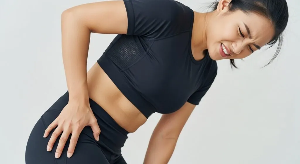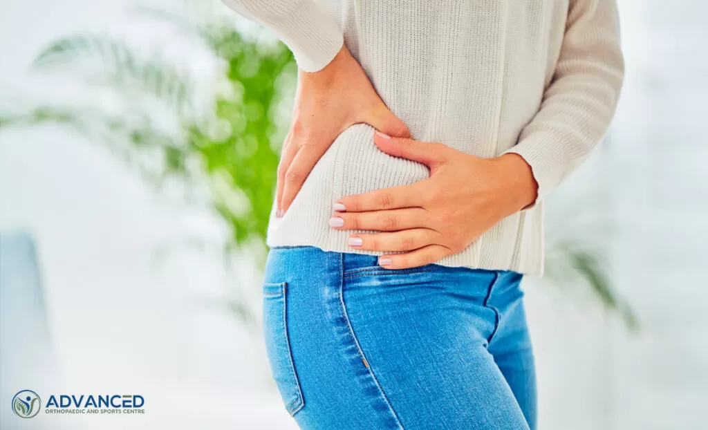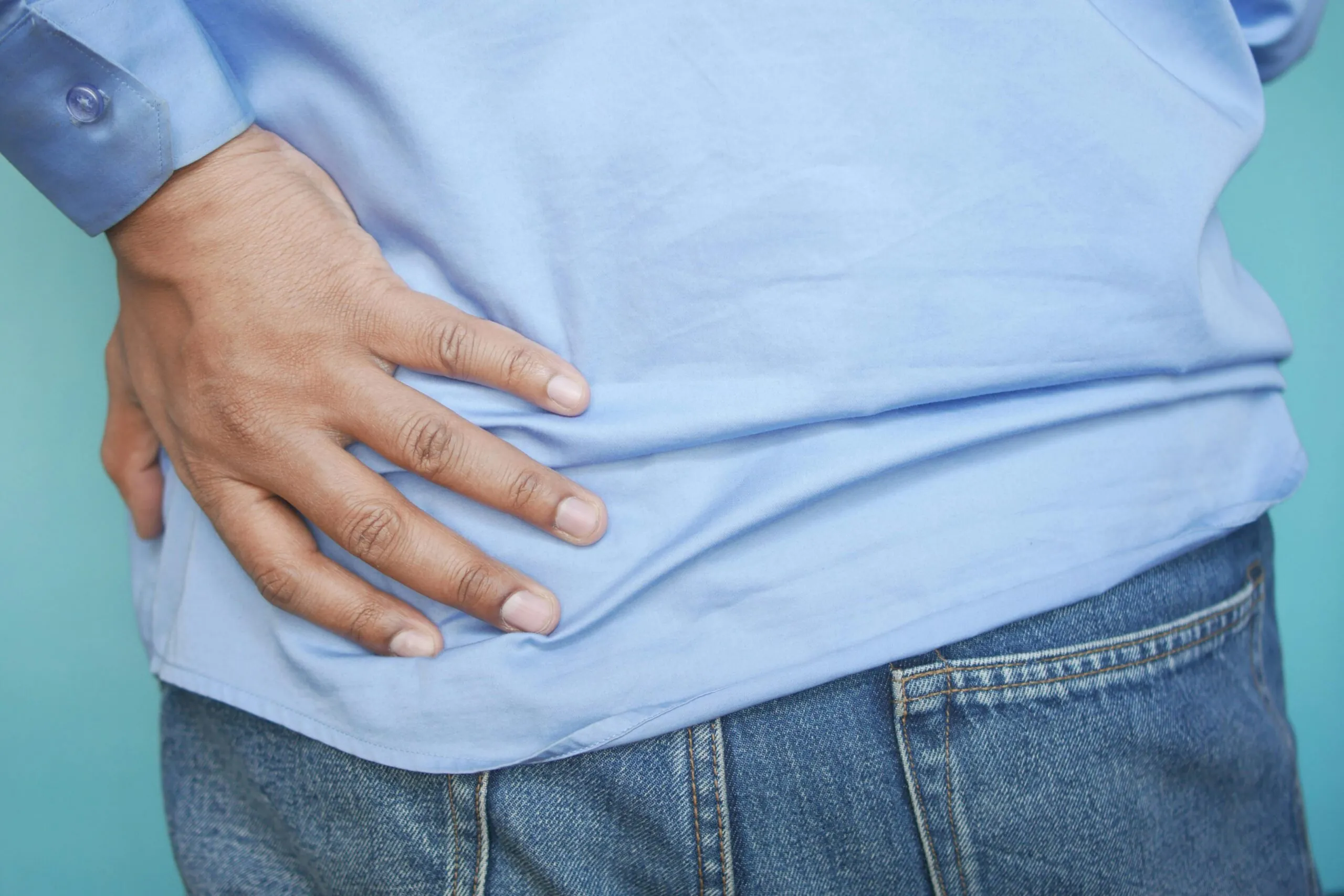
Did you know that climbing stairs can be more mechanically demanding on your hip tendons than running? Gluteal tendinopathy affects the tendons of the gluteus medius and minimus muscles where they attach to the greater trochanter, a bony prominence on the outer hip. It typically worsens with activities like climbing stairs, lying on the affected side, or prolonged standing on one leg.
The condition develops when repetitive loading exceeds the tendon’s capacity to repair and adapt. Walking mechanics, hip muscle strength imbalances, and habitual postures all influence how load is distributed through these tendons during daily activities. Accurate diagnosis is important because several conditions can produce similar symptoms. Treatment requires addressing the underlying tendon pathology rather than simply managing pain.
Anatomy and Function of the Gluteal Tendons
The gluteus medius and minimus muscles originate from the outer surface of the pelvis. They converge into tendons that insert onto the greater trochanter. The gluteal muscles act as the primary hip abductors, pulling the leg away from the body’s midline. Additionally, they function as external rotators, turning the leg outward. During single-leg stance, which occurs with every step of walking, they generate forces preventing the pelvis from dropping on the opposite side.
The gluteus medius tendon has distinct anterior, lateral, and posterior facets at its insertion. Each facet experiences different mechanical loads during movement. Moving the hip into adduction and internal rotation causes the iliotibial band to wrap across the gluteal tendon. This specific positioning creates significant compressive forces at the tendon’s anterior insertion.
This compression, combined with tensile loading during muscle contraction, creates a unique mechanical environment. Tendons generally tolerate tensile forces well but respond poorly to sustained compression. Activities and postures that repeatedly compress these tendons against bone contribute to tendon breakdown over time.
Recognising Gluteal Tendinopathy Symptoms
Pain localises to the lateral hip, typically within a hand’s width of the greater trochanter. Patients often describe a deep ache that intensifies with specific movements and positions rather than constant pain. The presentation includes:
- Difficulty lying on the affected side at night
- Pain climbing stairs or walking uphill
- Discomfort after sitting with legs crossed
Single-leg standing activities prove particularly provocative. Standing on the affected leg while dressing, getting out of a car, or shifting weight while cooking often reproduces symptoms. Prolonged walking, especially on uneven surfaces or inclines, gradually increases discomfort.
Morning stiffness commonly accompanies the condition. This distinguishes it from inflammatory arthritis (conditions like rheumatoid arthritis, in which the immune system attacks the joints), which typically causes prolonged morning stiffness. Pain may temporarily improve with gentle movement but worsens with continued loading or sustained positions.
Physical examination findings include:
- Tenderness directly over the greater trochanter
- Pain with resisted hip abduction
- Reproduction of symptoms during single-leg stance testing
The single-leg stance test, in which patients stand on the affected leg for a brief period, reliably reproduces lateral hip pain in patients with gluteal tendinopathy.
Contributing Factors and Risk Profiles
Load management issues represent the primary driver of gluteal tendinopathy. Sudden increases in walking distance, starting new exercise programmes, or returning to activity after periods of inactivity commonly precede symptom onset. The tendons require gradual adaptation to increased demands. Exceeding this adaptive capacity triggers the degenerative process.
Biomechanical factors influence tendon loading patterns. Hip adduction during gait—where the stance leg crosses towards the midline—increases compressive forces on the gluteal tendons. Weakness of the hip abductor muscles, deficits in trunk stability, and altered movement patterns after lower limb injuries all contribute to abnormal loading.
Postural habits play a role. Standing with weight shifted predominantly to one leg, sitting with legs crossed, or lying consistently on one side creates sustained compressive loads. These positions accumulate significant tendon stress over weeks and months.
Hormonal factors explain the higher prevalence in postmenopausal women. Oestrogen (a hormone that declines after menopause) influences tendon cell metabolism, collagen synthesis, and tissue hydration. The decline in oestrogen levels affects tendon structure and healing capacity. This makes tendons more vulnerable to repetitive loading injuries.
Diagnostic Assessment
Clinical examination forms the cornerstone of diagnosis. The combination of lateral hip tenderness, pain with resisted abduction in side-lying, and positive single-leg stance testing creates a reliable clinical picture. Symptoms localising specifically to the greater trochanter region, rather than the groin or buttock, help differentiate gluteal tendinopathy from hip joint or lumbar spine conditions.
Imaging confirms the diagnosis and assesses tendon condition. Ultrasound (a scan using sound waves to create images) visualises tendon thickening, altered echo texture, and any associated bursitis (inflammation of the fluid-filled cushioning sacs). MRI (magnetic resonance imaging, which uses magnetic fields to create detailed images of soft tissues) provides a detailed assessment of tendon structure. It detects partial-thickness tears and quantifies the extent of tendinopathic changes. These imaging tests help confirm the suspected condition and rule out other pathologies, including:
- Hip joint arthritis
- Stress fractures (tiny cracks in bone)
- Lumbar radiculopathy (nerve irritation in the lower back)
Healthcare providers assess the hip joint itself, as gluteal tendinopathy frequently coexists with hip osteoarthritis. Both conditions affect similar age groups and may share biomechanical contributors. Identifying concurrent hip joint pathology influences treatment planning and outcome expectations.
💡 Did You Know?
The gluteal tendons experience forces equivalent to several times body weight during single-leg activities. Running generates substantially higher tendon loads than walking.
Gluteal Tendinopathy Treatment: Conservative Management
Load management forms the foundation of conservative treatment. This involves temporarily reducing activities that provoke symptoms whilst maintaining general activity levels. Complete rest proves counterproductive. Tendons require mechanical loading to maintain structure and stimulate healing. A healthcare professional can help determine the appropriate load that stimulates adaptation without exceeding tissue tolerance.
Specific positions and movements warrant modification during the initial treatment phase. Avoiding crossing legs whilst sitting, distributing weight evenly when standing, and placing a pillow between knees when side-lying reduces compressive forces on the healing tendons. These adjustments often produce noticeable symptom reduction within the first few weeks.
Isometric exercises, or sustained muscle contractions without joint movement, provide the starting point for rehabilitation. Healthcare professionals typically recommend holding a gentle hip abduction contraction for a sustained period. You perform this several times daily. This loads the tendon within its current capacity whilst providing pain-relieving effects. Research demonstrates that isometric contractions reduce tendon pain for several hours following exercise.
Progressive loading exercises gradually increase tendon capacity over subsequent weeks and months. Standing hip abduction with resistance bands, single-leg balance progressions, and eventually sport-specific or activity-specific exercises systematically challenge the tendon. Symptom response guides progression. Exercises should not significantly increase pain during or after performance.
Physiotherapy and Exercise Rehabilitation
Structured physiotherapy programmes produce favourable outcomes. A physiotherapist assesses individual contributing factors such as muscle weakness patterns, movement quality, training history, and designs targeted interventions. This personalised approach addresses underlying deficits rather than applying generic protocols. A physiotherapist can establish exercise targets based on your specific condition, movement patterns, and recovery progress.
Hip abductor strengthening receives primary focus. However, rehabilitation extends beyond isolated muscle training. Trunk stability exercises improve pelvic control during single-leg activities. Hip external rotator and extensor strengthening addresses common concurrent weaknesses. The kinetic chain approach recognises that knee and ankle function influence hip mechanics.
Gait retraining benefits patients with identified walking pattern abnormalities. Increasing step width, reducing hip adduction during the stance phase, and modifying cadence alter the distribution of forces through the gluteal tendons. Real-time feedback, whether through mirrors or video, helps patients internalise new movement patterns.
Progression follows a staged approach over several months:
- Weeks 1-4: Pain management through load modification and isometric exercises
- Weeks 4-8: Isotonic strengthening in pain-free ranges, beginning functional exercises
- Weeks 8-12: Progressive resistance training, balance challenges, activity-specific rehabilitation
- Beyond 12 weeks: Return to full activity with ongoing maintenance exercises
⚠️ Important Note
Stretching the hip into adduction and internal rotation—common stretches targeting the iliotibial band or piriformis—compresses the gluteal tendons. This may aggravate symptoms. Avoid these positions during the rehabilitation period.
Injection Therapies
Corticosteroid injections (injections of anti-inflammatory medication) can provide short-term pain relief. However, they require careful consideration within the overall treatment plan. Healthcare providers direct the injection into the peritendinous space (the area around the tendon) or the associated bursa, rather than the tendon itself. Direct tendon injection risks weakening the tendon structure. Pain reduction creates a window for rehabilitation progression.
However, repeated corticosteroid injections carry risks. Studies demonstrate potential negative effects on tendon structure and healing capacity with multiple injections. The pain relief may encourage activity levels exceeding tendon tolerance. This potentially worsens the underlying condition. Current recommendations suggest limiting corticosteroid injections and combining them with structured rehabilitation.
Platelet-rich plasma (PRP) injections (a treatment that uses concentrated components of your own blood to promote healing) offer an alternative biological approach. PRP concentrates growth factors from the patient’s own blood. This theoretically enhances tendon healing responses. Research shows variable results.
Injection therapies work as adjuncts to rehabilitation rather than standalone treatments. The temporary pain relief enables patients to participate more effectively in exercise programmes that address underlying deficits and build tendon capacity.
Surgical Considerations
Healthcare providers reserve surgery for cases failing comprehensive conservative management over an extended period. The decision involves weighing symptom severity, functional limitations, and response to non-operative treatment against surgical risks and recovery requirements.
Surgical options include:
- Gluteal tendon repair (where the surgeon reattaches or repairs torn tendon tissue)
- Bursectomy (removal of the inflamed bursa)
- Iliotibial band release to reduce compressive forces
Endoscopic techniques, or minimally invasive procedures using small incisions and a camera, offer faster recovery times compared to open procedures.
Post-surgical rehabilitation follows similar principles to conservative management, with appropriate modifications to account for tissue-healing timelines. Protected weight-bearing progresses over the initial weeks. This is followed by gradual strengthening and return to function over several months. Full recovery from gluteal tendon surgery typically takes considerable time.
What Our Orthopaedic Surgeon Says
Patient education and active participation drive successful outcomes. Understanding why certain positions aggravate symptoms, how tendons adapt to loading, and what realistic recovery timelines look like, empowers patients to manage their rehabilitation.
The staged approach to loading deserves emphasis. Patients often feel frustrated by initial activity restrictions. However, this foundation allows subsequent progression without setbacks. Attempting to accelerate rehabilitation typically backfires. It triggers symptom flares that delay overall recovery.
Managing Daily Activities During Recovery
Modifying habitual positions reduces ongoing tendon compression. When standing for extended periods, distribute weight evenly between both legs rather than favouring one side. Use both handrails when climbing stairs to reduce single-leg loading. Choose chairs with armrests to assist with transitions between sitting and standing.
Sleep position adjustments help manage nighttime symptoms. If lying on the affected side, place a soft mattress topper or use a softer section of the mattress. When lying on the unaffected side, position a pillow between the knees to prevent the top leg from dropping into adduction. Lying on the back with a pillow under the knees provides an alternative comfortable position.
Footwear matters more than many patients realise. Supportive shoes with cushioned soles reduce ground reaction forces transmitted through the lower limb. Avoid flat, unsupportive shoes and high heels during the symptomatic period.
Pace daily activities to avoid accumulating excessive load. Breaking prolonged walking into shorter segments with rest periods improves symptom management compared with attempting extended distances. Planning rest days after higher-demand activities helps maintain progress.
When to Seek Professional Help
- Lateral hip pain persists beyond several weeks despite activity modification
- Night pain disrupting sleep despite position changes
- Increasing difficulty with stairs, walking, or getting in and out of vehicles
- Pain limits work duties or recreational activities
- Symptoms not responding to self-directed exercises
- Previous hip or lower limb injuries that may contribute to current symptoms
- Uncertainty about appropriate exercise progression
Commonly Asked Questions
How long does gluteal tendinopathy take to recover?
Recovery timelines vary based on individual factors. Tendons adapt slowly to loading. This requires patience with the progressive exercise approach.
Can I continue exercising with gluteal tendinopathy?
Yes, but activity modification is necessary. Low-impact exercises like swimming and cycling typically remain tolerable. Walking on flat surfaces in moderation usually continues. High-impact activities, prolonged single-leg exercises, and positions that provoke symptoms warrant temporary avoidance until tendon capacity improves.
What’s the difference between gluteal tendinopathy and trochanteric bursitis?
These conditions frequently coexist and produce similar symptoms. Trochanteric bursitis involves inflammation of the fluid-filled bursa overlying the greater trochanter. Gluteal tendinopathy affects the tendon structure itself. Current understanding recognises tendinopathy as the primary pathology in most cases, with bursitis occurring secondarily.
Will I need imaging to diagnose gluteal tendinopathy?
Clinical examination often provides sufficient diagnostic information to commence treatment. Imaging becomes valuable when symptoms don’t respond as expected, when excluding other pathology is important, or when assessing whether surgical intervention might be appropriate. Ultrasound (a scan using sound waves) and MRI (magnetic resonance imaging, which creates detailed images using magnetic fields) both visualise gluteal tendon pathology. Healthcare providers can use these as diagnostic tools to confirm the condition rather than as screening tests for healthy individuals.
Are cortisone injections helpful for gluteal tendinopathy treatment?
Corticosteroid injections (anti-inflammatory medication injected into the affected area) can provide short-term pain relief. This creates an opportunity for rehabilitation progression. However, they don’t address underlying tendon pathology. They may negatively affect tendon structure with repeated use. Injections work as an adjunct to comprehensive rehabilitation rather than a standalone treatment.
Please note: Individual recovery experiences will differ due to personal health factors. The information provided here is for general educational purposes. Consult qualified healthcare professionals for tailored advice specific to your condition rather than relying solely on this content.
Next Steps
Gluteal tendinopathy responds well to structured rehabilitation when load management and progressive strengthening are properly addressed. Early professional assessment identifies the specific biomechanical factors driving your symptoms and enables the development of a treatment plan tailored to your condition.
If you’re experiencing persistent lateral hip pain, difficulty climbing stairs, or pain when lying on your side, consult with an orthopaedic surgeon for a comprehensive evaluation and treatment planning.