Did you know that your hip pain today may stem from a structural problem that existed since birth? Hip dysplasia occurs when the hip socket fails to fully cover the femoral head (the thighbone), resulting in a joint that becomes unstable over time. The shallow socket concentrates pressure on a smaller surface area. This accelerates cartilage breakdown and leads to early-onset arthritis—sometimes decades before typical age-related joint wear appears.
Adults with hip dysplasia often adapt unconsciously to their condition. They develop compensatory movement patterns that mask the underlying problem. The hip may initially feel “different” rather than painful. It may have subtle clicking during certain movements or fatigue after walking.
How Hip Dysplasia Develops in Adults
Hip dysplasia exists on a spectrum from mild to severe socket undercoverage. Some individuals are born with borderline anatomy that functions adequately during childhood and young adulthood. It only becomes symptomatic when activity levels increase or the body’s compensatory mechanisms fail. Others have developmental dysplasia that was either missed during infant screening or treated incompletely.
The hip joint relies on congruent surfaces to distribute load evenly across thick cartilage. In dysplasia, the socket edge bears disproportionate stress. This causes the labrum (the ring of fibrocartilage, a tough but flexible tissue that lines the socket rim) to work harder, stabilising the joint. This overloaded labrum frequently tears. It often serves as the first symptom-producing event in an otherwise stable but abnormal hip.
Hormonal changes, particularly during pregnancy, can unmask previously asymptomatic dysplasia. Relaxin hormone loosens pelvic ligaments. This removes compensatory stability and reveals underlying joint inadequacy.
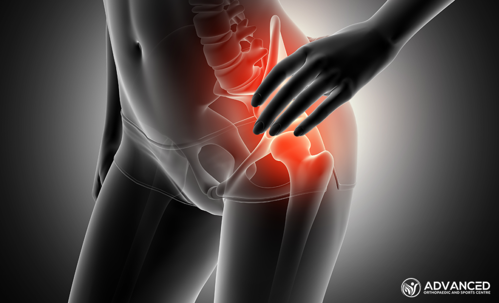
Recognising Hip Dysplasia Symptoms
Groin Pain Pattern
Hip dysplasia pain localises to the anterior groin crease. It is often described as deep within the joint. This differs from lateral hip pain, which typically indicates bursitis (inflammation of fluid-filled cushions near the joint) or tendon problems. Patients frequently demonstrate the “C-sign,” cupping their hand around the front of the hip to indicate the site of pain.
Pain typically worsens with prolonged sitting, especially in low chairs or car seats that flex the hip beyond a certain angle. Rising from a seated position elicits sharp catching sensations. Walking on uneven surfaces or climbing stairs increases discomfort. Unlike muscle strains, which typically improve with rest, dysplastic hip pain often worsens after periods of inactivity.
Movement Limitations
Internal rotation—turning the knee inward while keeping the hip flexed—becomes restricted early in the progression of dysplasia. Patients may notice difficulty crossing legs, putting on shoes, or entering and exiting vehicles. These limitations often develop so gradually that individuals attribute them to ageing or inflexibility rather than joint pathology.
The affected hip may also demonstrate a clicking or clunking sensation during specific movements. This particularly happens when transitioning from sitting to standing. While not all hip clicks indicate problems, reproducible catching accompanied by pain warrants investigation.
Compensatory Changes
Years of adapting to an unstable hip produce visible changes in posture and gait. The pelvis may tilt forward to increase the coverage of the socket. This creates an exaggerated lumbar curve and secondary lower back pain. The affected leg often externally rotates during walking, giving a slightly waddling appearance.
Muscle imbalances develop as certain groups overwork while others weaken. Hip flexors (muscles that lift your thigh) become tight from constant protective activity. Gluteal muscles (the buttock muscles that stabilise the hip) lose strength from altered biomechanics. These changes perpetuate symptoms even after addressing the primary joint problem.
Diagnostic Approach
Clinical Examination
Orthopaedic assessment begins with observing gait patterns and standing posture. Specific provocative tests stress the hip joint in ways that reproduce dysplasia-related symptoms. The anterior impingement test—a manoeuvre in which the physician moves the hip through specific positions (bending, bringing it across the body, and rotating it inward)—typically produces groin pain when labral damage is present.
Range-of-motion measurement quantifies restrictions, particularly by comparing internal rotation between sides. Strength testing identifies muscle imbalances that require rehabilitation, regardless of surgical decisions.
Imaging Studies
Plain radiographs (X-rays) remain fundamental for diagnosing hip dysplasia. Specific measurements quantify socket coverage and joint congruency. The lateral centre-edge angle measures how far the socket extends over the femoral head. Lower angles indicate dysplasia, while borderline angles represent borderline coverage.
MRI with contrast injection (arthrogram)—in which a contrast agent is injected into the joint to enhance visibility of structures on the scan—visualises soft-tissue damage that is not visible on X-rays. Labral tears, cartilage defects, and early degenerative changes guide treatment planning. The pattern and extent of damage influence whether joint-preserving surgery remains viable.
CT scanning with three-dimensional reconstruction precisely maps anatomy for surgical planning. Understanding the exact shape of the socket deficiency allows surgeons to plan corrective procedures accurately.
Treatment Options for Hip Dysplasia in Adults Singapore
Conservative Management
Activity modification reduces symptoms without altering disease progression. Avoiding deep hip flexion (bending the hip far forward), prolonged sitting, and high-impact activities decreases joint stress. Swimming and cycling typically prove better tolerated than running or court sports.
Physiotherapy addresses modifiable factors contributing to symptoms. Strengthening hip stabilisers—particularly the gluteus medius (a buttock muscle that helps maintain pelvic level) and the deep external rotators (small muscles that rotate the hip outward)—improves dynamic joint control. Manual therapy techniques address muscle tightness and joint mobility restrictions. While physiotherapy cannot correct anatomical abnormalities, optimising surrounding structures often provides meaningful symptom relief.
Anti-inflammatory medications reduce pain associated with synovitis (inflammation of the joint lining) and cartilage damage. Cortisone injections (medication delivered directly into the joint to reduce inflammation) provide temporary relief. This relief is useful for confirming the hip as the source of pain or for managing symptoms while planning treatment.
💡 Did You Know?
The labrum contributes to hip stability by creating a seal that maintains negative pressure within the joint. It effectively acts like a suction cup that helps hold the femoral head in place.
Joint-Preserving Surgery
Periacetabular osteotomy (PAO)—a procedure where the surgeon cuts and repositions the hip socket bone to better cover the ball of the hip joint—improves femoral head coverage. This procedure reorients the existing bone rather than replacing it. It can help preserve the natural joint when performed before significant arthritis develops. Candidates have painful dysplasia with preserved cartilage and sufficient bone stock for reconstruction. Your surgeon will determine whether this option is appropriate for your specific hip anatomy and overall health.
Recovery from PAO requires several months of protected weight-bearing (using crutches or limiting how much weight you put on the leg) followed by intensive rehabilitation. High-impact activities may remain inadvisable. Outcomes vary among patients based on individual health factors.
Arthroscopic surgery (minimally invasive surgery using a small camera and instruments inserted through tiny incisions) addresses labral tears and removes damaged cartilage fragments. In dysplastic hips, arthroscopy serves as an adjunct to osteotomy or a palliative measure when joint-preserving reconstruction isn’t appropriate. Arthroscopy alone, without correcting underlying bony abnormality, provides limited long-term benefit for dysplasia.
Joint Replacement
Hip replacement may become necessary when arthritis progresses beyond joint-preserving options. Dysplastic hips present technical challenges. Shallow sockets require special techniques to achieve stable implant fixation. Altered anatomy demands careful surgical planning.
Modern implants and surgical approaches can achieve outcomes even in challenging dysplastic anatomy. Conservative management and timely joint-preserving surgery can significantly delay this.
Living with Hip Dysplasia
Activity Considerations
Understanding which activities load the hip excessively allows informed lifestyle choices. Single-leg stance—present during stair climbing, running, and standing on one leg—significantly increases the body-weight force through the hip. Reducing these activities decreases joint stress without eliminating exercise entirely.
Water-based activities support body weight while allowing cardiovascular conditioning and muscle strengthening. Cycling provides appropriate exercise with minimal hip impact when the seat height prevents excessive flexion. Yoga and Pilates can improve core stability and flexibility. They require modification of poses involving deep hip flexion or extreme rotation.
Weight Management
Body weight translates to considerable force through the hip during walking. Weight reduction is a non-surgical intervention for hip pain of any cause.
Monitoring Progression
Regular clinical and radiographic assessment tracks disease progression. This enables timely intervention. Annual X-rays reveal cartilage space narrowing or bony changes indicating advancing arthritis. Increasing symptoms despite adequate conservative measures prompt reassessment of surgical options.
Preparing for Your Orthopaedic Consultation
Documenting symptoms prior to the appointment improves consultation efficiency. Note what activities worsen pain, where exactly discomfort localises, and how symptoms have changed over time. Including prior imaging studies enables comparison with current findings.
Consider your goals and concerns regarding treatment. Understanding whether you wish to avoid surgery, return to specific activities, or address pain interfering with daily life helps your surgeon tailor recommendations to your individual needs and circumstances.
Prepare questions regarding expected outcomes, recovery timelines, and the risks associated with various treatment approaches. Understanding the natural history of untreated dysplasia and the outcomes of intervention enables informed decision-making.
When to Seek Professional Help
- Groin or anterior hip pain lasting more than several weeks
- Clicking or catching sensation accompanied by pain
- Progressive difficulty with activities previously performed easily
- Pain that awakens you from sleep or persists at rest
- Visible limping or gait changes noticed by others
- Lower back or knee pain that hasn’t responded to direct treatment
Commonly Asked Questions
Can hip dysplasia appear for the first time in adults?
Hip dysplasia doesn’t develop in adulthood; the abnormal anatomy is present from birth or early childhood. However, symptoms often first appear in adults when compensatory mechanisms fail, activity demands exceed joint capacity, or hormonal changes during pregnancy reveal underlying instability.
Will exercise make hip dysplasia worse?
Appropriate exercise helps manage hip dysplasia by strengthening stabilising muscles and maintaining mobility. However, high-impact activities like running and jumping may accelerate cartilage wear in abnormal joints. Low-impact conditioning through swimming, cycling, and targeted strengthening supports the joint while maintaining fitness.
How quickly does hip dysplasia progress to arthritis?
Progression varies among patients based on individual health factors. It depends on the severity of dysplasia, activity levels, body weight, and other factors. Regular monitoring identifies progression requiring intervention. Your doctor will track your specific condition to recommend treatment at the appropriate time.
Is hip dysplasia hereditary?
Hip dysplasia has genetic components. If you have hip dysplasia, screening family members, particularly female relatives, may identify individuals who would benefit from monitoring or early intervention.
Can I still exercise after periacetabular osteotomy?
Most patients with PAO can return to active lifestyles, including recreational sports. High-impact activities like running may remain inadvisable to protect the reconstructed joint. Swimming, cycling, hiking, and many other activities typically become possible after complete recovery. Response times vary depending on your specific condition and rehabilitation compliance.
Individual recovery experiences and treatment outcomes vary based on personal health factors. This content provides general educational information and should not replace personalised medical advice. Please consult qualified healthcare professionals for guidance tailored to your specific situation.
Next Steps
Early evaluation of hip pain—particularly groin discomfort in younger adults—allows identification of hip dysplasia before significant arthritis develops. Joint-preserving surgery can offer better outcomes when performed while cartilage remains healthy. Accurate diagnosis enables treatment planning that preserves long-term joint function.
If you’re experiencing groin pain, hip clicking, or difficulty with activities like putting on shoes or climbing stairs, an orthopaedic surgeon can evaluate whether hip dysplasia is the cause and discuss treatment options.