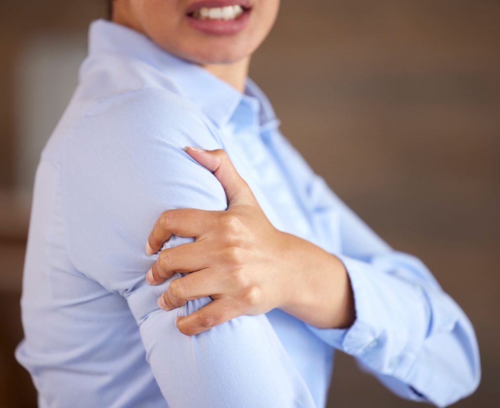
The shoulder joint features a wide range of motion, which can sometimes influence its baseline structural stability. As one of the body’s highly flexible joints, it relies extensively on surrounding soft tissue structures alongside its native bony contours to stay in place. When these structures weaken or sustain damage, the humeral head (ball) can potentially shift within the glenoid (socket), creating an unsettling sensation that your shoulder might slip out of position.
This feeling typically differs from general shoulder pain. Shoulder instability symptoms can include a sense of looseness, apprehension during certain movements, and sometimes a noticeable joint shift. This sensation may become more apparent with overhead activities, reaching behind the back, or during sports involving throwing or physical contact.
The underlying cause, whether a traumatic injury, repetitive stress, or inherent tissue laxity, helps determine the presentation of symptoms and guides the most appropriate management strategy.
Anatomy of Shoulder Stability
Multiple structures work in coordination to help keep your shoulder joint aligned: the labrum, joint capsule, rotator cuff muscles, and supporting ligaments.
The labrum is a ring of fibrocartilage attached to the rim of the glenoid socket. It aims to deepen the naturally shallow socket cavity and serves as a structural anchor point for the shoulder ligaments. This configuration increases the contact area between the ball and socket, supporting the joint’s baseline stability.
The joint capsule surrounds the shoulder joint like a protective sleeve. Within this capsule, thickened bands form the glenohumeral ligaments, which typically tighten at different arm positions to help manage translation. The inferior glenohumeral ligament complex is an important component involved in restricting excessive forward displacement when the arm is positioned in abduction and external rotation, a position commonly reached during the cocking phase of throwing.
The rotator cuff comprises four muscles designed to compress the humeral head into the socket during movement. These muscles provide dynamic stability and active muscular control that complements the passive restraints of the ligaments and labrum. When rotator cuff strength or coordination diminishes, the shoulder can lose a portion of this compressive force and may become more susceptible to abnormal shifting.
Types of Shoulder Instability
Traumatic Instability
A single forceful event—typically a fall onto an outstretched arm, a direct blow to the shoulder, or a forced external rotation—can tear the labrum and stretch or rupture supporting ligaments. First-time traumatic dislocations can cause structural damage that may increase the risk of developing recurrent instability over time.
The common injury pattern involves an anterior (forward) dislocation associated with a Bankart lesion, where the labrum detaches from the front of the socket. Posterior dislocations occur less frequently, occasionally resulting from a fall onto an outstretched arm, direct trauma, or specific medical events. Recurrent dislocation episodes can potentially cause additional damage to already compromised joint structures.
Atraumatic Instability
Some individuals develop joint looseness without a history of specific trauma. Generalised joint hypermobility, often referred to as being naturally flexible or “double-jointed,” is characterised by inherently loose shoulder capsules. Engaging in repetitive overhead activities, such as swimming, volleyball, or tennis, can gradually stretch ligaments. Atraumatic instability typically affects both shoulders and may involve excessive movement in multiple directions (multidirectional instability), with symptoms usually developing gradually rather than suddenly.
Microtraumatic Instability
Athletes performing repetitive overhead motions can develop instability through accumulated minor injuries. Each throwing motion or swimming stroke creates small stresses on capsular structures. Over time, these microtraumas accumulate, resulting in a stretched capsule that no longer provides adequate restraint.
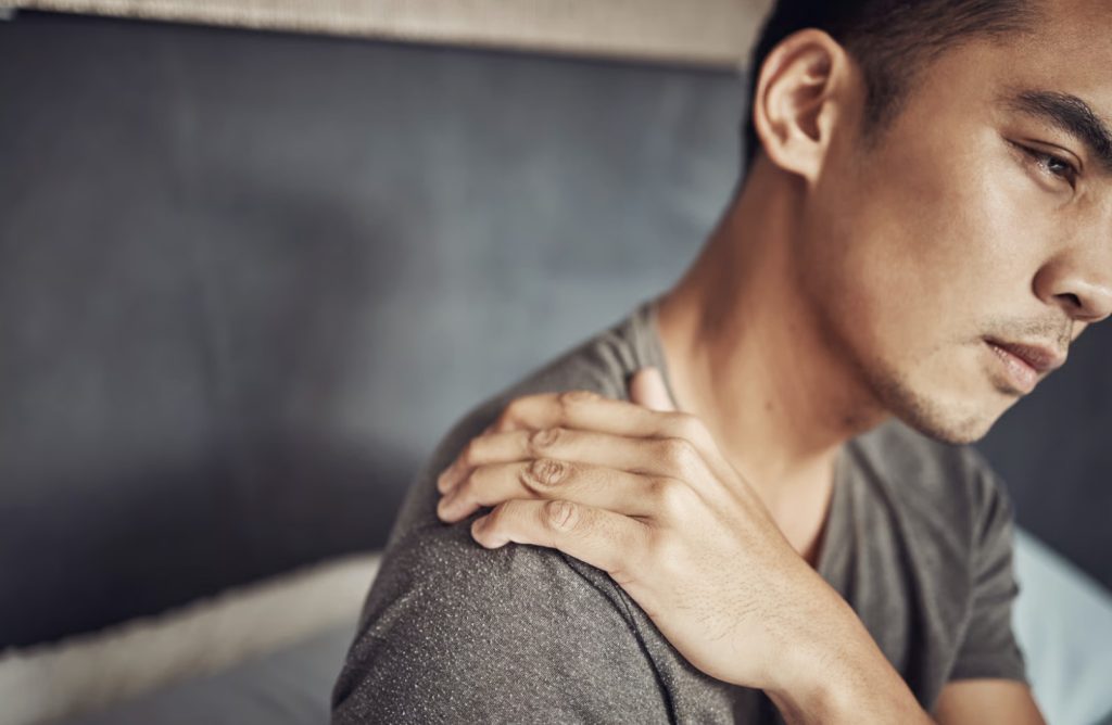
Recognising Shoulder Instability Symptoms
The hallmark symptom is apprehension, an uncomfortable sense that the shoulder will dislocate during specific movements. This feeling intensifies when the arm moves into vulnerable positions, particularly when raised to shoulder height and rotated outward.
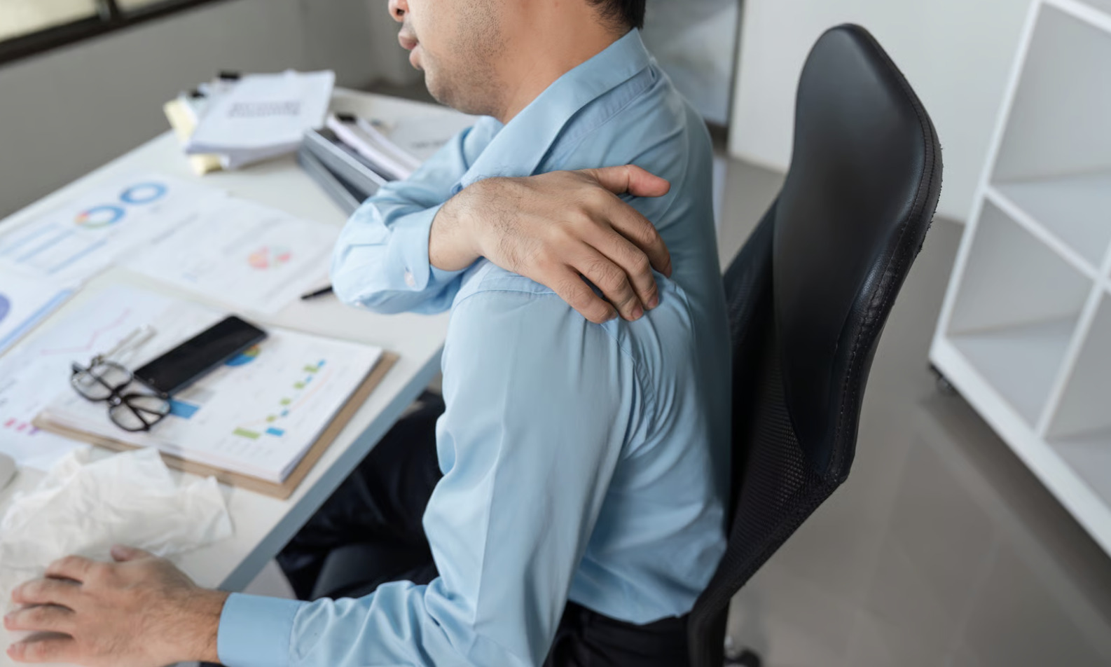
Physical sensations include:
- A feeling of looseness or “dead arm” during activities
- Clicking, catching, or grinding within the joint
- Sensation of the shoulder shifting or sliding
- Sudden sharp pain with certain arm positions
- Weakness when lifting objects away from the body
Functional limitations often involve:
- Difficulty sleeping on the affected side
- Avoidance of overhead reaching or lifting
- Inability to throw with previous force or accuracy
- Hesitation during activities requiring arm elevation
- Compensatory movements using the opposite arm
Shoulder instability symptoms may be subtle initially. Many people unconsciously modify their activities to avoid triggering positions, masking the true extent of dysfunction until the condition progresses.
Did You Know?
The shoulder socket covers only about one-third of the humeral head surface, comparable to a golf ball sitting on a tee. This anatomical arrangement prioritises range of motion but requires intact soft tissues for stability.
Common Causes and Risk Factors
- Previous dislocation is considered a significant predictor of future joint looseness. Observations suggest that the younger an individual is at the time of their first dislocation, the higher the likelihood of recurrence is, as structural damage from an initial event may not always fully resolve without targeted intervention.
- Sports participation can also influence overall structural risk. Contact sports (such as rugby or football) can expose the shoulder to direct trauma, while overhead sports (such as tennis, swimming, or cricket) can create repetitive stress on stabilising structures.
- Anatomical variations can affect baseline stability parameters. A shallower glenoid socket, a smaller labrum profile, or a naturally loose capsule provides less inherent constraint. These variations can become clinically relevant when combined with demanding physical activities.
- Connective tissue characteristics influence how ligaments behave under load. Conditions affecting collagen, such as Ehlers-Danlos syndrome, are associated with systemic joint laxity. Even without a diagnosed condition, some individuals naturally possess more elastic soft tissues.
- Muscle weakness or imbalance can compound other underlying risk factors. Inadequate rotator cuff strength reduces the efficiency of dynamic stabilisation, while scapular muscle dysfunction can alter overall shoulder mechanics, potentially placing greater demands on passive soft tissue structures.
How Orthopaedic Specialists Evaluate Shoulder Instability
Clinical evaluation typically begins with a detailed history-taking. The mechanism of any past injuries, the frequency of episodes of looseness, specific positions that trigger symptoms, and the overall impact on daily activities help guide the assessment.
Physical examination includes targeted clinical manoeuvres designed to evaluate instability symptoms under controlled conditions. The apprehension test involves positioning the arm in a vulnerable orientation while the examiner notes the patient’s muscle response and comfort level. The load-and-shift test directly assesses the degree of humeral head translation within the socket cavity.
Imaging studies provide essential structural data:
- X-rays help identify associated bony changes, such as fractures of the glenoid rim or indentation fractures of the humeral head (Hill-Sachs lesion) that can occur during a displacement.
- MRI visualises soft tissue structures, helping to identify potential damage to the labrum, capsule, or rotator cuff.
- MR arthrography, utilising a joint contrast injection, aims to enhance the detection of deep labral tears.
- CT scanning allows for precise measurements of bone architecture if surgical planning requires detailed visualisation of bone loss.
The combination of clinical findings and imaging observations is generally utilised to evaluate the nature of the instability and help guide appropriate management options.
Non-Surgical Management Approaches
Physiotherapy forms the foundation of initial treatment for most presentations of instability. A structured programme focuses on:
- Strengthening rotator cuff muscles to improve dynamic stability
- Scapular stabilisation exercises to optimise shoulder blade positioning
- Proprioceptive training to enhance joint position awareness
- Sport-specific rehabilitation for athletes
Treatment typically lasts several months, with gradual improvement in strength and control. Compliance with home exercises significantly influences outcomes.
Activity modification reduces exposure to destabilising forces during recovery. This may involve temporarily avoiding overhead sports, adjusting training intensity, or making technique corrections that place less stress on the shoulder.
Bracing provides external support during activities. Shoulder stabilisation braces limit movement into vulnerable positions while allowing functional use. Bracing works best as an adjunct to rehabilitation rather than a standalone treatment.
Important Note
Shoulder instability that follows a significant traumatic dislocation, particularly in younger or highly active individuals, is often closely evaluated for surgical stabilisation options to help manage the risk of recurrence. Discussing appropriate care timelines with a professional can be helpful, as repeated instability events can potentially increase the risk of progressive structural changes over time.
Surgical Treatment Options
When conservative management does not restore adequate joint stability or when structural tissue damage is substantial, surgical alternatives may be considered to address the underlying pathology.
- Arthroscopic Stabilisation: This technique utilises small incisions and a specialised camera to evaluate and repair damaged structures from within the joint space. Surgeons aim to reattach the torn labrum to the glenoid rim using small anchors and sutures, and a capsular plication may be performed to tighten stretched ligaments. Arthroscopic approaches are generally associated with a customised recovery protocol and the management of post-operative discomfort.
- Open Stabilisation: This approach provides direct visualisation for complex cases. An open Bankart repair may be considered when significant glenoid rim bone loss is present or when previous arthroscopic interventions have not fully resolved the looseness.
- Bone Augmentation Procedures: These operations address more extensive bone deficiencies. The Latarjet procedure transfers a small portion of the coracoid bone, along with its attached muscle, to the anterior glenoid, aiming to reinforce the baseline bony contours and provide dynamic muscular support.
Post-surgical rehabilitation follows a structured physical therapy protocol spanning several months, with a gradual return to full activity typically discussed between four and six months, depending on the specific procedure and individual healing rates.
Preventing Recurrence and Progression
Maintaining shoulder stability requires ongoing attention to the factors that protect the joint.
- Consistent strengthening of the rotator cuff and scapular muscles preserves dynamic stability. These exercises should continue indefinitely, not just during symptomatic periods.
- Proper technique in sports and daily activities reduces unnecessary stress on stabilising structures. Coaching input helps athletes identify and correct movement patterns that increase the risk of injury.
- Gradual return to activity after any episode of instability allows tissues to adapt to increasing demands. Rushing back to full participation before adequate healing and strengthening increases the likelihood of recurrence.
- Recognition of warning signs enables early intervention. Increasing episodes of instability, worsening apprehension, or declining function warrant reassessment even if initial treatment seemed successful.
When to Seek Professional Help
- The shoulder has dislocated or feels like it is partially slipping out of place.
- Persistent sensation of looseness that affects daily activities
- Inability to perform work or sport requirements due to shoulder instability
- Pain or apprehension when moving the arm into certain positions
- Previous instability treatment that no longer controls symptoms
- Weakness or “dead arm” episodes during throwing or lifting
Commonly Asked Questions
Can shoulder instability heal on its own?
Mild looseness stemming from temporary muscle weakness or minor ligament stretching may improve with a dedicated, structured rehabilitation program. However, structural changes such as complete labral tears or significant ligament disruptions typically do not spontaneously return to their original baseline state. The body may form scar tissue, but it can lack the structural alignment of original tissues, occasionally leaving residual joint looseness.
How do I know if my shoulder actually dislocated?
Complete dislocation causes obvious deformity, severe pain, and inability to move the arm. The shoulder appears squared-off rather than rounded. Many people describe a sensation of the shoulder “going out and coming back in,” which represents subluxation (partial dislocation) rather than complete dislocation, though both indicate instability.
Will I need to stop playing sports if I have shoulder instability?
Treatment goals include a return to desired activities. Many athletes successfully return to sport after appropriate rehabilitation or surgery. The approach depends on the sport’s demands, the severity of instability, and individual goals. Overhead and contact athletes may require more intensive treatment to achieve reliable stability.
How long does recovery take after shoulder stabilisation surgery?
Initial healing requires immobilisation in a sling for several weeks. Physiotherapy progresses through phases over four to six months. Return to non-contact activities typically occurs around three to four months, while contact sports or heavy overhead work may require six months or longer.
Can shoulder instability affect both shoulders?
Atraumatic instability from generalised laxity commonly affects both shoulders. Traumatic instability in one shoulder does not directly cause problems in the other shoulder, though similar injury mechanisms could affect either shoulder. Hypermobile individuals should be particularly attentive to both shoulders.
Next Steps
Structural changes affecting the labrum or ligament components generally do not resolve spontaneously, and successive instability events can potentially impact the surrounding joint tissues over time. Early medical evaluation, whether concluding in a customised physiotherapy protocol or a discussion of surgical stabilisation alternatives, helps establish an appropriate care plan before further structural changes develop. An orthopaedic assessment aims to identify the specific anatomical factors driving your symptoms to help determine a suitable course of care.
If you are experiencing shoulder looseness, apprehension when raising or rotating your arm, or episodes in which your joint feels as if it shifts out of position, consulting an orthopaedic surgeon in Singapore can provide a comprehensive evaluation to safely review your treatment options.