An Achilles tendon rupture is a tear in the strong fibrous cord that connects the calf muscles to the heel bone. This tendon plays a key part in walking, running, and jumping by enabling the foot to push off the ground. A rupture can happen suddenly during sports or daily activities, making movement difficult and requiring medical evaluation. This guide covers the symptoms, causes, types, diagnosis, and treatment options for an Achilles tendon rupture.
Symptoms of an Achilles Tendon Rupture
An Achilles tendon rupture often causes immediate and noticeable symptoms. Recognising these signs early can help determine when to seek medical attention.
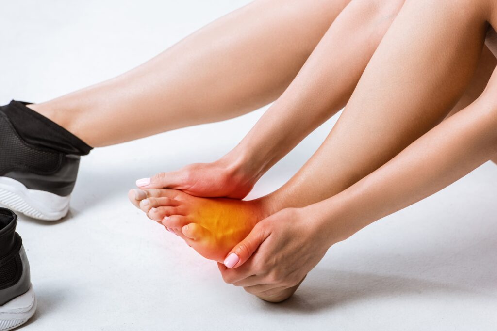
Sharp Pain at the Back of the Leg: A sudden, intense pain occurs in the lower leg or heel, often described as feeling like a direct impact. In some cases, discomfort may be mild, particularly in individuals with pre-existing tendon conditions.
A Popping Sound or Sensation: Many people hear or feel a “pop” at the time of injury, followed by immediate weakness in the leg.
Loss of Strength and Stability: The foot may feel weak or unsteady, making it difficult to push off while walking, climb stairs, or stand on tiptoes.
Limited Ability to Point the Toes Downward: Movement of the foot becomes restricted, particularly the ability to press down or lift the heel off the ground.
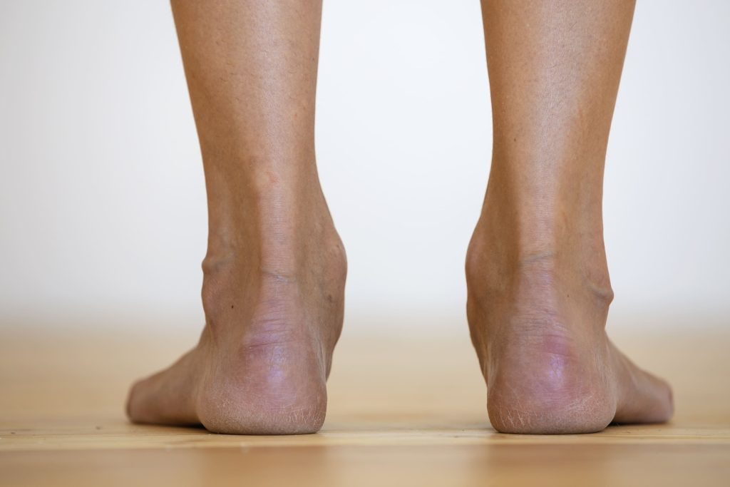
Visible or Palpable Gap in the Tendon: A small gap or indentation may be felt 2–6 cm above the heel bone, where the tendon has torn.
Swelling and Bruising: The affected area becomes swollen, tender to the touch, and may develop bruising over the following hours or days.
What Causes an Achilles Tendon Rupture?
An Achilles tendon rupture occurs when the tendon is overstretched beyond its capacity. This can happen due to sudden force, underlying tendon wear, or other contributing factors.
Sudden High-Impact Movements: Rapid acceleration or abrupt stops in sports like football, basketball, and tennis place extreme stress on the Achilles tendon, increasing the risk of rupture.
Natural Changes with Age: The tendon gradually loses elasticity over time, making individuals over 30 more prone to injury, especially during strenuous activity.
Previous Tendon Problems: A history of Achilles tendinitis, chronic inflammation, or previous minor tears weakens the tendon and raises the likelihood of a rupture.
Use of Certain Medications: Fluoroquinolone antibiotics and corticosteroid treatments (both oral and injected) have been linked to tendon weakness, making ruptures more likely.
Underlying Medical Conditions: Conditions such as rheumatoid arthritis, diabetes, and gout can affect tendon strength, increasing the risk of injury.
How Is an Achilles Tendon Rupture Diagnosed?
A thorough clinical examination and imaging tests help confirm an Achilles tendon rupture and guide treatment planning.
Physical Examination: The doctor gently squeezes the calf while the patient lies face down with their feet hanging off the edge of the table. If the foot does not move in response, it indicates a complete rupture.
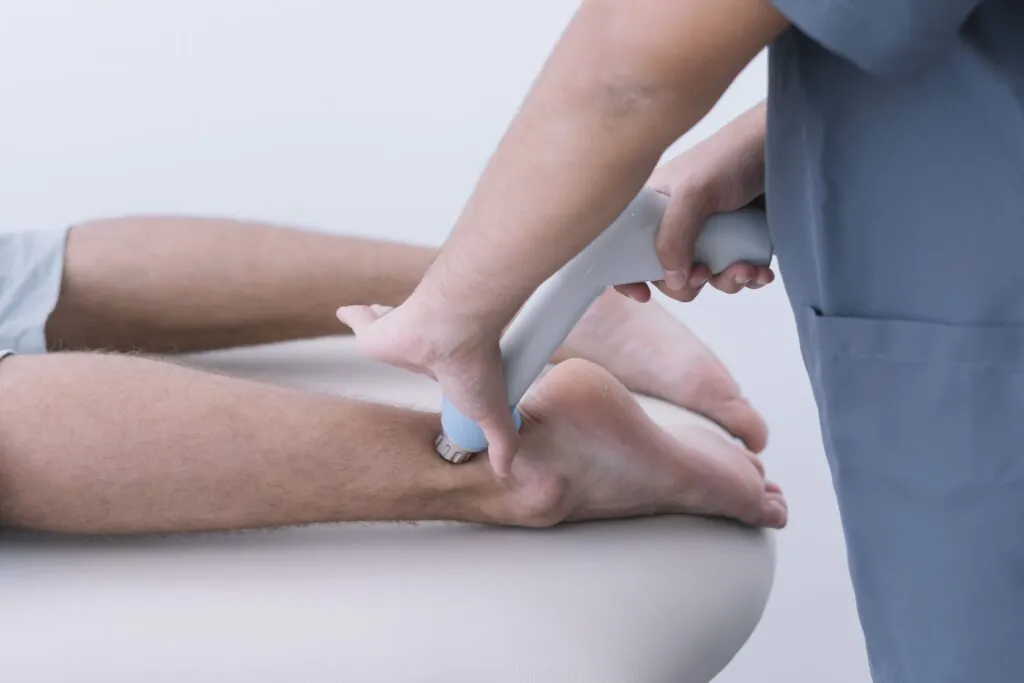
Ultrasound Imaging: High-frequency sound waves create real-time images of the tendon, showing the location and severity of the tear. This test also assesses tendon movement when the foot is flexed.
MRI Scan: This provides a detailed view of the tendon structure and surrounding tissues, helping determine whether surgery is needed. It is particularly useful for evaluating chronic ruptures or complex injuries.
X-rays: Although X-rays do not show soft tissue injuries, they help rule out fractures or other bone-related conditions that may cause similar symptoms.
Treatment Options for an Achilles Tendon Rupture
The approach to treating an Achilles tendon rupture depends on factors such as the severity of the tear, the patient’s age, overall health, and activity level. Both surgical and non-surgical options aim to restore tendon function and mobility while minimising the risk of re-injury.
Non-Surgical Treatment
Non-surgical treatment is often recommended for individuals with partial ruptures or those with lower activity levels. The goal is to allow the tendon to heal naturally while maintaining stability and preventing excessive movement.
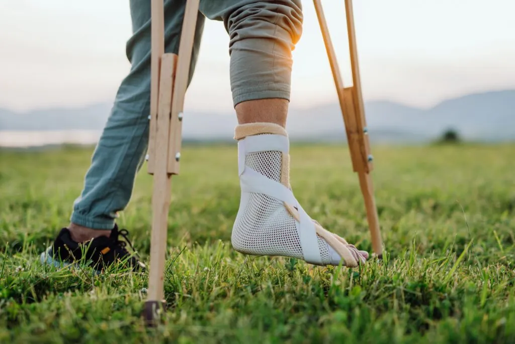
Immobilisation with a Boot or Cast: The foot is placed in a downward (plantarflexed) position using a walking boot or cast, which keeps the torn tendon ends close together. Over 6–8 weeks, the angle of the boot is gradually adjusted to encourage proper healing while preventing excessive strain. During this period, weight-bearing is typically introduced in stages based on medical advice.
Rehabilitation Programme: After immobilisation, a structured physiotherapy programme is introduced to restore mobility, rebuild strength, and prevent stiffness. Initial exercises focus on gentle range-of-motion movements before progressing to strengthening activities. Full recovery typically takes 4–6 months, with improvements continuing beyond this timeframe.
Platelet-Rich Plasma (PRP) Injections: Some practitioners use PRP therapy, where a concentrated solution of the patient’s platelets is injected into the tendon to support healing. However, research on its effectiveness remains inconclusive, and it is generally considered a supplementary treatment rather than a primary option.
Surgical Treatment
Surgical repair is often recommended for complete ruptures, particularly in younger or more active individuals. Surgery can reduce the likelihood of re-rupture and may allow a more structured recovery compared to non-surgical approaches.
Open Repair: A single incision is made over the rupture site, and the torn tendon ends are sutured together. This approach allows direct visualisation of the tendon, ensuring a strong repair, but it carries a slightly higher risk of wound complications such as infection or delayed healing.
Minimally Invasive Repair: Small incisions are used instead of a large open incision, allowing sutures to be passed through the tendon with less disruption to surrounding tissue. This technique reduces the risk of wound-related issues while still providing secure tendon repair.
Augmented Repair for Chronic Ruptures: If the rupture remains untreated for several weeks or the tendon ends have retracted, additional techniques may be required. Tendon grafts or transfers from nearby muscles can reinforce the repair and restore function. Recovery is usually longer and involves an extended rehabilitation plan to regain mobility and strength.
Preventing Achilles Tendon Injuries
Minimising strain on the Achilles tendon can reduce the risk of rupture and support long-term tendon health. Stretching, strength training, and controlled activity progression help maintain flexibility and resilience. Gradual increases in exercise intensity lower the chance of injury, particularly in high-impact activities like sprinting and jumping. Supportive footwear and training on suitable surfaces can reduce excessive tendon stress. Those with a history of Achilles tendinitis should monitor symptoms closely and address any discomfort early to prevent further damage.
Conclusion
An Achilles tendon rupture can limit movement, but recovery is achievable with the appropriate treatment. Whether treated surgically or non-surgically, following a structured rehabilitation plan helps restore function and strength. Early medical assessment and a tailored recovery approach can support better long-term outcomes.
For advice on treatment options and recovery, schedule a consultation today to take the next step towards restoring strength and mobility.