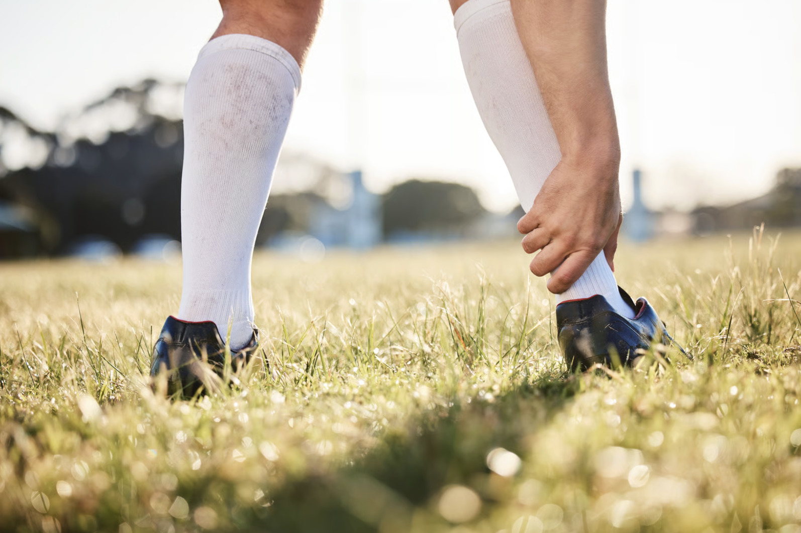
A distinct “pop” at the back of your ankle during a sprint or jump is one of the most reliable signs that your Achilles tendon has ruptured—not simply become inflamed. The Achilles tendon connects your calf muscles to your heel bone and transmits substantial forces during running. This mechanical demand makes it vulnerable to two distinct injuries: tendonitis (inflammation and microdamage) and rupture (partial or complete tear). These conditions require different treatment approaches and have vastly different recovery timelines.
Tendonitis develops gradually through repetitive stress. Ruptures typically occur suddenly during explosive movements (such as jumping, sprinting, or sudden changes in direction). Both cause pain in the same anatomical location. Their onset patterns, pain characteristics, and functional limitations differ significantly.
The distinction between Achilles tendon rupture vs tendonitis extends beyond pain levels. It determines whether conservative treatment will suffice or surgical intervention becomes necessary.
How Achilles Tendonitis Develops
Achilles tendonitis results from cumulative microtrauma (repeated small-scale injuries) that exceeds the tendon’s repair capacity. The condition progresses through predictable stages. Each stage has distinct characteristics.
Reactive Tendonitis
Initial inflammation occurs when training load increases too rapidly. The tendon thickens as cells proliferate in response to stress. Pain appears during warm-up. It often diminishes mid-activity before returning afterward. Morning stiffness lasting less than 30 minutes is typical. The tendon may feel tender to touch but maintains normal strength.
Degenerative Tendinopathy
Prolonged overuse leads to structural changes within the tendon. Collagen fibres (the protein structures that give the tendon its strength) become disorganised. The tendon loses its normal elastic properties. Pain becomes more persistent. It occurs during daily activities like climbing stairs. The tendon may develop nodular thickening palpable along its length, typically 2-6 centimetres above the heel bone insertion.
Insertional vs Mid-Portion Tendonitis
Insertional tendonitis affects the tendon’s attachment to the heel bone. It is often associated with bone spurs and bursitis (inflammation of fluid-filled cushioning sacs). Mid-portion tendonitis occurs in the watershed zone where blood supply is poorest. Treatment approaches differ: insertional cases may require heel modifications, while mid-portion cases respond better to eccentric loading exercises (exercises where the muscle lengthens under tension).
Recognising an Achilles Tendon Rupture
Complete or partial Achilles ruptures present differently from tendonitis. The injury mechanism and immediate symptoms provide strong diagnostic clues.
The Rupture Moment
Many people describe hearing or feeling a distinct “pop” or “snap” at the back of the ankle. The sensation resembles being kicked or struck, though no external contact occurred. This moment marks the actual tendon failure. Immediate pain varies. Some experience severe discomfort whilst others report surprisingly little pain after the initial injury.
Immediate Physical Signs
A visible gap or depression may appear in the tendon, typically 2-6 centimetres above the heel. Swelling develops rapidly, often within the first hour. Bruising follows, sometimes extending down to the foot or up the calf. The calf muscle may appear bunched higher than usual as it retracts without the tendon anchoring it.
Functional Impairment
Walking becomes immediately difficult or impossible. Attempting to rise onto tiptoes fails completely with full ruptures. Pushing off during walking feels weak or absent. Climbing stairs requires pulling with handrails rather than pushing with the affected leg.
The Thompson Test: A Simple Self-Assessment
The Thompson test (also called the calf squeeze test) provides useful preliminary information about tendon integrity. Whilst not definitive, it helps determine urgency for professional evaluation.
Performing the Test
- Lie face down on a bed with your feet hanging over the edge
- Have someone firmly squeeze your calf muscle midway between the knee and ankle
- Observe your foot’s response
Interpreting Results
A normal response shows the foot pointing downward (plantarflexion) when the calf is squeezed. Absence of foot movement suggests the tendon may be torn. Weak or minimal movement may indicate partial tear. Comparing both legs helps identify subtle differences in response strength.
💡 Did You Know?
The Thompson test works because squeezing the calf muscle transmits force through the intact Achilles tendon, pulling the heel bone and pointing the foot downward. Without tendon continuity, this mechanical chain breaks.
Comparing Symptoms: Rupture vs Tendonitis
Pain Onset and Character
Tendonitis pain builds gradually over weeks to months. It worsens with increased activity. Rupture pain occurs instantly during a specific movement—jumping, sprinting, or sudden direction change. Tendonitis creates an aching, burning sensation. Rupture produces sharp, localised pain that may quickly subside.
Activity Tolerance
With tendonitis, exercise remains possible though uncomfortable. Warm-up often temporarily reduces symptoms. With rupture, weight-bearing activities become severely limited immediately. No amount of warming up restores function.
Physical Examination Findings
Tendonitis shows diffuse tenderness along the tendon, possible thickening, and preserved strength (though painful). Rupture demonstrates a palpable gap, positive Thompson test, and inability to perform single-leg heel raise.
Risk Factors for Each Condition
Tendonitis Risk Factors
- Rapid increases in training volume or intensity strain the tendon’s adaptive capacity
- Tight calf muscles reduce tendon flexibility
- Inappropriate footwear fails to support proper biomechanics
- Running on hard surfaces increases impact loading
- Age-related tendon changes reduce resilience, with tendonitis frequently seen between ages 30-50
Rupture Risk Factors
- Previous tendonitis weakens the tendon structure, increasing rupture vulnerability
- Fluoroquinolone antibiotics (a class of antibiotics including ciprofloxacin and levofloxacin) and corticosteroid injections (anti-inflammatory medications injected directly into the area) may compromise tendon integrity
- Systemic conditions including diabetes and rheumatoid arthritis affect tendon health
- Sporadic high-intensity activity without regular conditioning—the “weekend warrior” pattern—creates vulnerability
- Ruptures frequently occur between ages 30-40 in active individuals
⚠️ Important Note
If you’ve received corticosteroid injections into or around the Achilles tendon, or have taken fluoroquinolone antibiotics, inform your orthopaedic specialist. These medications alter tendon properties and influence treatment decisions.
Diagnostic Approaches
Clinical examination provides substantial diagnostic information. Imaging confirms the diagnosis and characterises injury severity.
Physical Examination
Your orthopaedic specialist will assess gait pattern, tendon continuity, strength testing, and range of motion. The Thompson test forms part of standard evaluation. Palpation (feeling the area with their hands) identifies tenderness location, gaps, or nodular changes. Single-leg heel raise testing quantifies functional capacity.
Ultrasound Imaging
Ultrasound visualises tendon structure in real-time using sound waves to create images. It shows thickening, tears, or complete discontinuity. Dynamic examination assesses tendon movement during muscle contraction. This accessible, non-invasive modality provides immediate results. It allows comparison with the opposite side.
MRI Evaluation
MRI (magnetic resonance imaging) offers detailed cross-sectional images created using magnetic fields and radio waves. It distinguishes partial from complete tears. Tendonitis appears as signal changes within the tendon substance. Ruptures show fluid-filled gaps and retracted tendon ends. MRI helps surgical planning by measuring gap distance and assessing tissue quality.
Treatment Pathways for Tendonitis
Conservative management (non-surgical treatment approaches) successfully resolves most tendonitis cases. Treatment addresses both symptoms and underlying mechanical factors.
Load Management
Reducing aggravating activities allows tissue healing whilst maintaining fitness through cross-training (alternative exercises that don’t stress the Achilles tendon). Complete rest is counterproductive. Tendons require appropriate loading to heal properly. Gradual return to activity follows pain response. Increase load only when symptoms remain stable.
Eccentric Exercise Programme
The most widely studied protocol involves twice-daily sessions over approximately 12 weeks, performing sets with both straight and bent knee. Some studies suggest that a lower-volume “do-as-tolerated” approach over a similar period may produce comparable results, reducing the burden of the full repetition count. Discomfort during exercises is generally expected and acceptable; significant increases in pain may indicate excessive loading and should prompt reassessment.
Adjunct Treatments
- Heel lifts temporarily reduce tendon strain
- Appropriate footwear provides cushioning and support
- Night splints maintain tendon length
- Shockwave therapy (a treatment using sound wave pulses to stimulate healing) may benefit cases unresponsive to exercise alone
- Platelet-rich plasma injections (injections using concentrated components from your own blood to promote healing) remain under investigation with variable evidence
Treatment Options for Ruptures
Achilles ruptures require prompt treatment decisions. Both surgical and non-surgical approaches can achieve good outcomes when appropriately selected.
Surgical Repair
When surgery is indicated, it is ideally performed within the first two weeks of injury for optimal tissue quality and repair conditions. Patients who present later can still be assessed for surgical or conservative management, though your surgeon will advise on the most appropriate approach based on the delay, tissue condition, and your individual circumstances.
Non-Surgical Management
Functional rehabilitation uses a walking boot with progressive heel-wedge reduction, achieving healing through scar tissue formation. This approach avoids surgical risks and can be comparable to surgical results when initiated early with strict protocol adherence. Patient selection and compliance significantly influence success.
Factors Influencing Treatment Choice
Activity level and functional demands are important considerations. Surgical repair may be associated with lower re-rupture rates, though it also carries higher risks of complications including wound infection and nerve injury. Conservative management with early functional rehabilitation can achieve comparable functional outcomes in many patients. Higher-demand patients should discuss both approaches with their surgeon, as individual factors — including timing of presentation, overall health, and personal preference — all contribute to shared decision-making.
Recovery Timeline Expectations
Tendonitis Recovery
Mild tendonitis may resolve within a few months of appropriate management. Chronic tendonitis with degenerative changes requires several months of consistent rehabilitation. Some cases need longer recovery, particularly when symptoms have persisted for years before treatment. Return to sport follows graduated protocols based on functional testing.
Rupture Recovery
Initial immobilisation or protected weight-bearing typically spans 6 to 8 weeks regardless of treatment approach. Progressive strengthening follows, advancing to functional exercises over the subsequent months. Return to jogging typically occurs around 4 to 6 months, with full return to competitive sport generally requiring 9 to 12 months — though individual timelines vary based on injury severity, treatment approach, and rehabilitation commitment.
✅ Quick Tip
Track your symptoms daily during recovery. Note pain levels, activities performed, and any setbacks. This record helps your orthopaedic specialist fine-tune your rehabilitation programme.
What Our Orthopaedic Surgeon Says
Distinguishing between tendonitis and rupture often becomes clear through careful history-taking. The mechanism of injury—gradual onset versus sudden event—provides useful diagnostic direction. However, partial ruptures can masquerade as severe tendonitis. This is why imaging plays an important role when clinical findings are ambiguous.
Early intervention may improve outcomes for both conditions. For tendonitis, addressing the problem before degenerative changes become established may shorten recovery. For ruptures, initiating treatment within the first two weeks—whether surgical or conservative—supports healing potential.
Putting This Into Practice
- Apply immediate RICE protocol for acute injuries: rest from aggravating activities, ice for 15-20 minutes several times daily, compression with bandage support, and elevation above heart level
- Document your injury pattern including when symptoms started, what triggered them, and how they’ve progressed. Note any audible pop, visible changes, or functional limitations
- Modify activities appropriately by avoiding hills, stairs, and pushing movements that stress the tendon. Use supportive footwear and consider temporary heel lifts
- Seek timely professional evaluation especially if you experienced sudden-onset symptoms, cannot perform heel raises, or notice a gap in the tendon
When to Seek Professional Help
- Sudden pain with a popping sensation during activity
- Inability to rise onto tiptoes or push off when walking
- Visible gap or depression in the tendon
- Rapid swelling and bruising around the ankle
- Persistent tendon pain unresponsive to two weeks of rest
- Morning stiffness lasting more than 30 minutes
- Pain that worsens despite activity modification
- Previous Achilles problems with new or changing symptoms
Commonly Asked Questions
Can an Achilles tendon rupture heal without surgery?
Complete ruptures can heal through functional rehabilitation protocols using a boot with graduated heel wedges. Scar tissue bridges the gap, restoring continuity. This approach avoids the risks associated with surgery — including wound infection, nerve injury, and other complications — which are meaningfully higher with operative management. Re-rupture rates may be somewhat higher with conservative treatment than with surgery, though evidence suggests the difference is smaller than once thought when early functional rehabilitation is used. Success depends on timely initiation, strict protocol adherence, and appropriate patient selection. Your doctor can advise which approach is most suitable based on your specific situation.
How can I tell if my Achilles pain is serious?
Indicators that warrant prompt evaluation include sudden onset during activity, a popping sensation, inability to perform single-leg heel raises, visible deformity or gap in the tendon, and rapid swelling. Tendonitis symptoms that fail to improve with rest, worsen progressively, or significantly limit daily activities also warrant professional evaluation to prevent progression.
Will my Achilles tendonitis become a rupture?
Chronic tendonitis creates structural changes that increase rupture risk. Progression isn’t inevitable. Proper management through load modification, eccentric exercises, and addressing biomechanical factors allows tendon healing and reduces vulnerability. Continuing high-level activity despite persistent symptoms increases progression risk.
How long should I wait before seeing a doctor for Achilles pain?
Sudden-onset symptoms suggesting rupture require evaluation within days to ensure timely treatment initiation. For gradual-onset tendonitis symptoms, trying activity modification and ice for one to two weeks is reasonable. Seek evaluation sooner if symptoms worsen, significantly limit function, or don’t respond to initial self-care measures.
Can I still walk with a ruptured Achilles tendon?
Limited walking remains possible using compensatory muscle activation. Gait appears abnormal and pushing off feels markedly weak. Some individuals don’t realise they’ve sustained a rupture. They attribute symptoms to a severe strain. However, continued walking on an untreated rupture can increase the gap between tendon ends. This potentially complicates later repair.
General Disclaimer: Individual recovery experiences and treatment outcomes will differ based on personal health factors, injury severity, activity level, and adherence to treatment protocols. The information provided is for educational purposes and should not replace consultation with qualified healthcare professionals. Always consult your orthopaedic specialist or healthcare provider for personalised advice tailored to your specific medical situation and health profile.
Next Steps
Injury mechanism and the Thompson test result are the two most important factors in distinguishing a rupture from tendonitis. For tendonitis, eccentric loading exercises and graduated return to activity form the basis of treatment. For ruptures, treatment—whether surgical or conservative—should begin within two weeks of injury to support optimal healing.
If you are experiencing sudden-onset Achilles pain with a popping sensation, inability to rise onto tiptoes, or a visible gap in the tendon, consult a foot and ankle specialist in Singapore promptly. Persistent tendon symptoms unresponsive to two weeks of rest also warrant evaluation.


