Knee surgery addresses structural problems that conservative treatments cannot resolve. Modern surgical techniques range from minimally invasive arthroscopy to total joint replacement, each targeting specific conditions and damage patterns. The choice depends on your age, activity level, extent of damage, and which knee structures need repair.
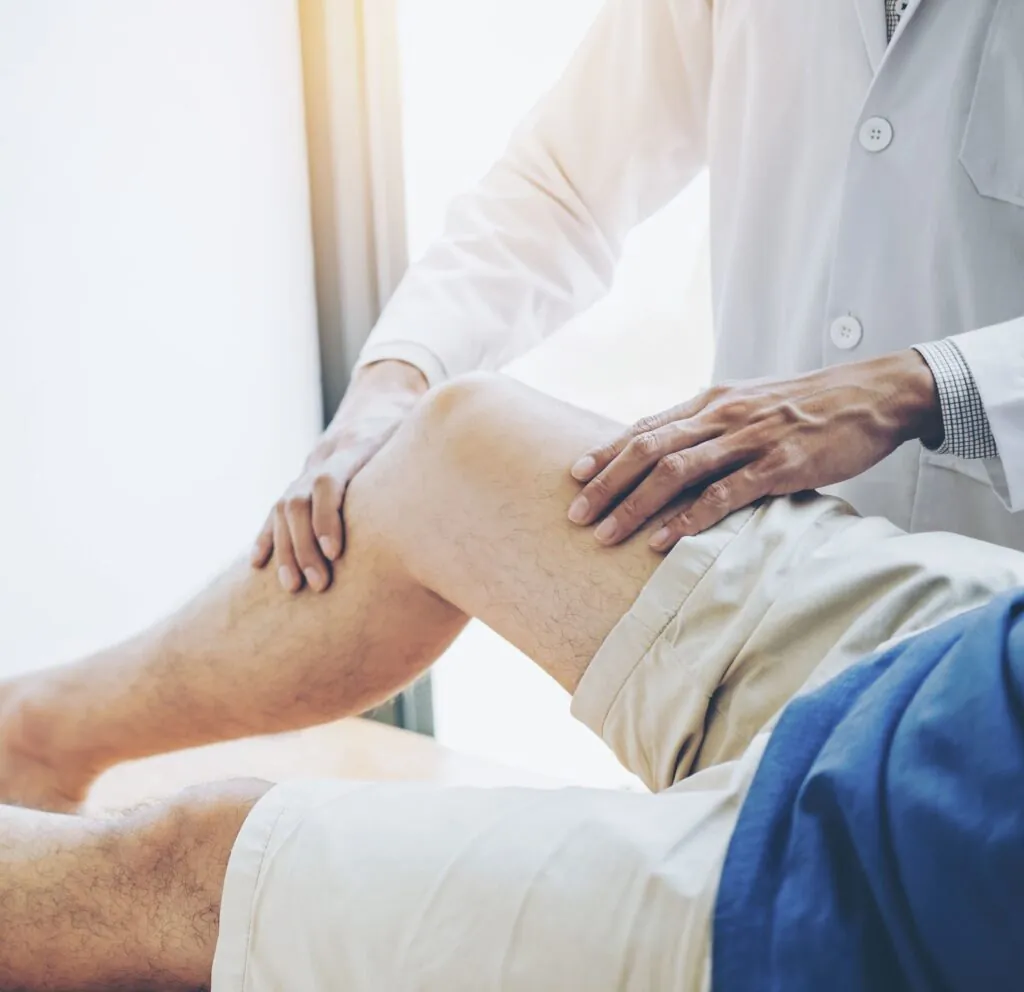
Orthopaedic surgeons evaluate multiple factors before recommending surgery: cartilage wear patterns, ligament integrity, bone alignment, and inflammatory markers. Imaging studies, including X-rays, MRI scans, and sometimes CT scans, reveal the precise location and severity of damage. This comprehensive assessment determines whether you need tissue repair, joint preservation, or replacement surgery.
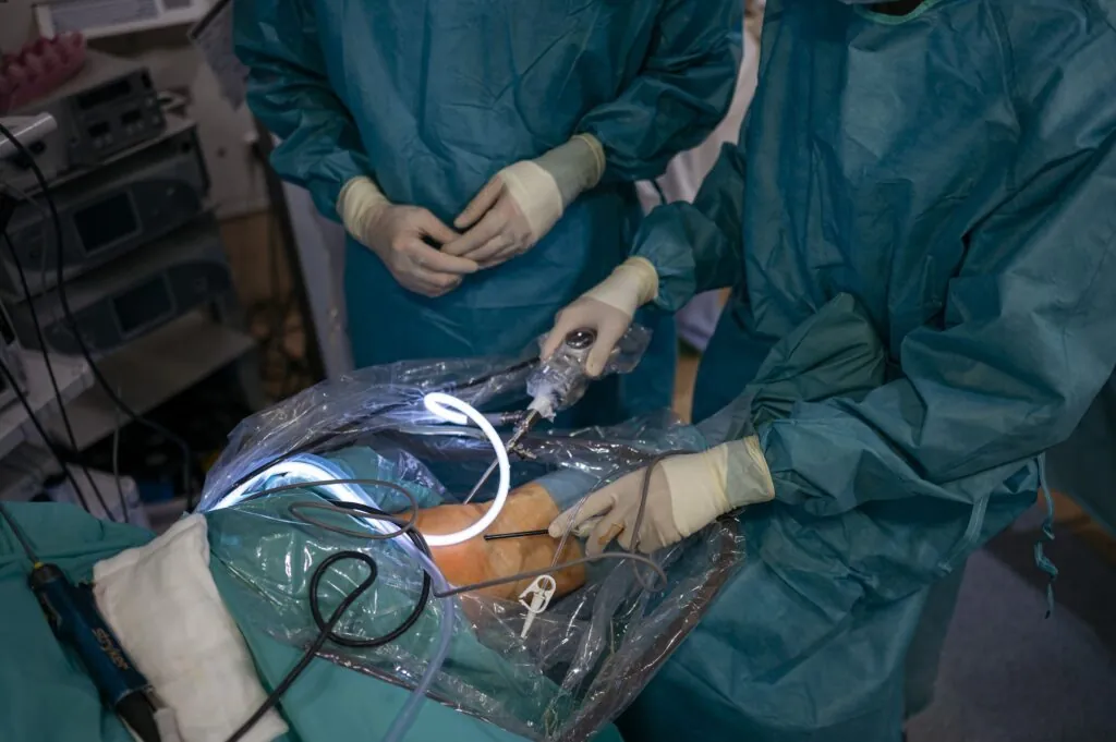
Arthroscopic Surgery
Arthroscopy uses pencil-sized instruments and a tiny camera inserted through small incisions around your knee. Surgeons visualise and treat internal joint problems while minimising tissue disruption. Recovery typically takes 2-6 weeks, depending on the procedures performed.
Standard arthroscopic procedures include:
Meniscus Repair or Removal
Torn meniscus fragments cause catching, locking, and persistent pain. Surgeons either stitch tears in the outer blood-rich zone or remove damaged portions that cannot heal. Meniscus preservation techniques work well for younger patients with fresh tears near the meniscus edge.
Loose Body Removal
Cartilage or bone fragments floating within the joint space trigger sudden pain and mechanical symptoms. Arthroscopic removal provides immediate relief and prevents further cartilage damage from these fragments grinding against joint surfaces.
Synovectomy
Inflamed synovial tissue in conditions like rheumatoid arthritis produces excess fluid and inflammatory substances. Removing this tissue reduces pain and swelling, though the condition may require ongoing medical management.
Microfracture and Drilling
Small holes created in exposed bone stimulate bleeding and healing responses. This technique works for focal cartilage defects smaller than 2 square centimetres, particularly in younger patients with isolated injuries.
Cartilage Restoration Procedures
Cartilage damage creates persistent pain and limits function. Several surgical techniques address cartilage defects based on size, location, and patient factors.
Autologous Chondrocyte Implantation (ACI)
ACI harvests your own cartilage cells during arthroscopy, grows them in a laboratory for 3-5 weeks, then reimplants millions of cells into the defect. This two-stage procedure is suitable for larger defects (2-10 square centimetres) in younger, active patients.
The cultured cells mature into hyaline-like cartilage over 12-18 months. Rehabilitation follows strict protocols: non-weight-bearing for 6 weeks, progressive loading over months, and avoidance of impact activities for 1 year.
Osteochondral Autograft Transfer (OATS)
OATS procedures transplant healthy cartilage and underlying bone from non-weight-bearing knee areas to damaged regions. Cylindrical plugs fill defects immediately with mature cartilage. Single or multiple plugs reconstruct areas up to 4 square centimetres.
Recovery progresses faster than ACI since the transplanted cartilage functions immediately. Patients use crutches for 4-6 weeks, then gradually return to activities over 4-6 months. Donor site healing rarely causes problems when harvest is performed using proper techniques.
Osteochondral Allograft Transplantation
Fresh donor cartilage and bone are used to treat larger defects or multiple-compartment damage. Grafts come from tissue banks within 28 days of harvest to maintain cell viability. Precise sizing and surgical technique ensure proper fit and healing.
Immunosuppression isn’t required since cartilage lacks blood vessels. Grafts take 3-4 months to incorporate with careful rehabilitation. Long-term success depends on graft integration and patient compliance with activity modifications.
Realignment Osteotomy
Osteotomy corrects abnormal knee alignment, causing uneven wear. Surgeons cut and reposition the tibia or femur, shifting weight from damaged to healthy cartilage areas. This joint-preserving surgery is suitable for younger patients with single-compartment arthritis and malalignment.
High Tibial Osteotomy
Bow-legged alignment overloads the inner knee compartment. Opening or closing wedge techniques realign the tibia, transferring forces to the healthier outer compartment. Modern plates and surgical guides improve precision and outcomes.
Bone healing takes 3-4 months with progressive weight bearing. Full recovery requires 6-12 months. Athletes often return to sports after complete healing.
Distal Femoral Osteotomy
Knock-knee deformity damages the outer compartment. Femoral cuts above the knee restore neutral alignment. This technically demanding procedure requires careful planning and execution.
Recovery mirrors that of tibial osteotomy, with similar timelines. Combined procedures address complex deformities affecting multiple planes. Computer navigation and patient-specific guides enhance accuracy.
Partial Knee Replacement
💡 Did You Know?
Partial knee replacements preserve all cruciate ligaments and most of the joint, allowing more natural knee movement patterns compared to total replacement. The retained tissues provide better proprioception and stability.
Minimally invasive techniques use 3-4 inch incisions. Hospital stays average 1-2 days with faster recovery than total replacement. Patients walk immediately and resume normal activities within 6-8 weeks. The range of motion typically exceeds total knee replacement.
Long-term success requires appropriate patient selection. Obesity, inflammatory arthritis, and ligament damage predict poorer outcomes. Revision to total replacement remains straightforward if arthritis progresses to other compartments.
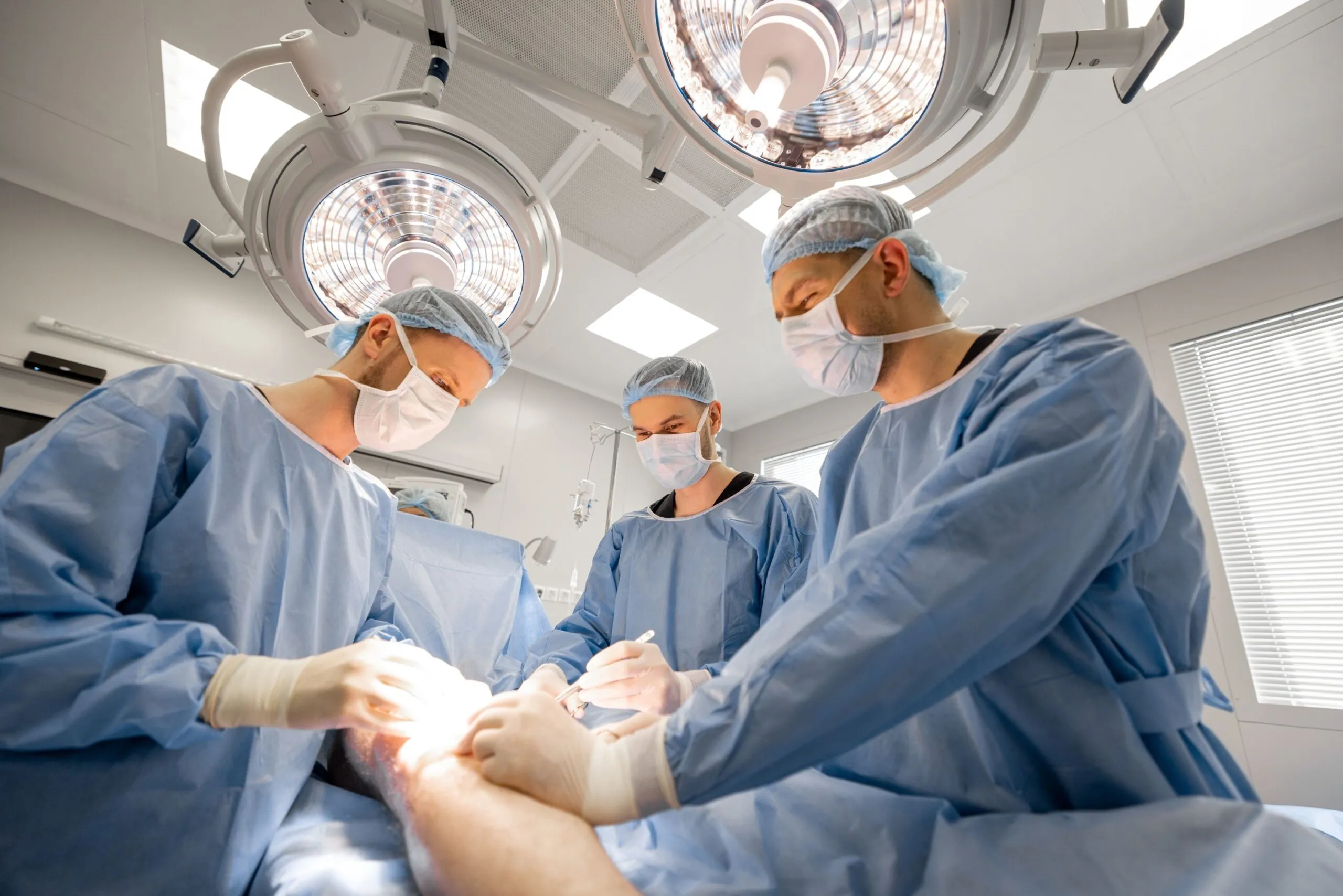
Total Knee Replacement
Total knee arthroplasty resurfaces all three compartments with metal and plastic components. Modern implants replicate natural knee geometry while providing smooth, pain-free motion. Surgeons remove minimal bone, preparing surfaces to accept precisely sized components.
Surgical Technique
Incisions measure 6-8 inches along the front of your knee. Surgeons remove damaged cartilage and small amounts of underlying bone. Trial components verify proper sizing, alignment, and stability before cementing final implants.
Current techniques include:
- Computer navigation for precise alignment
- Patient-specific instruments based on MRI or CT scans
- Robotic assistance for bone preparation
- Cementless fixation in younger patients
Recovery Timeline
Hospital discharge occurs 1-3 days post-surgery. Physical therapy begins immediately with walking, range-of-motion exercises, and strength training. Most patients walk without assistive devices by 4-6 weeks.
Recovery milestones:
- Week 1-2: Walking with a walker or crutches
- Week 3-4: Transition to cane, driving (automatic transmission)
- Week 6-8: Walking without aids, returning to light work
- Month 3: Most daily activities are comfortable
- Month 6-12: Final improvements in strength and endurance
Implant Longevity
Modern knee replacements function well beyond 20 years in most patients. Polyethene wear, loosening, and infection represent primary failure modes. Younger, heavier, and more active patients face higher risks of revision.
Activity modifications protect implants: avoiding repetitive impact, maintaining a healthy weight, and choosing low-impact exercises. Regular follow-up detects problems early, when simpler revision options are available.
Ligament Reconstruction
Anterior cruciate ligament (ACL) tears cause instability during pivoting activities. Reconstruction uses tendon grafts to create a new ligament, restoring stability for active lifestyles.
Graft Options
Patellar Tendon Autograft
The central third of your patellar tendon with bone blocks provides strong initial fixation. Bone-to-bone healing occurs by 6-8 weeks. Kneeling discomfort may persist, but most athletes return to cutting sports successfully.
Hamstring Autograft
Semitendinosus and gracilis tendons create a four-strand graft without disturbing the extensor mechanism. Harvest site morbidity remains minimal. Fixation methods continue to improve, with outcomes comparable to those of the patellar tendon.
Quadriceps Tendon Autograft
Partial-thickness quadriceps tendon, with or without bone, provides robust tissue for revision cases or larger patients. This option preserves hamstrings for athletes requiring sprinting power.
Allograft
Donor tissue eliminates harvest morbidity but requires longer incorporation. Younger patients show higher failure rates with allografts. Older recreational athletes benefit from shorter surgery and easier initial recovery.
⚠️ Important Note
ACL reconstruction doesn’t guarantee return to previous activity levels. Success depends on surgical technique, graft choice, rehabilitation compliance, and psychological readiness. Second ACL injuries occur more frequently than initial tears.
What Our Orthopaedic Surgeon Says
Surgical timing significantly impacts outcomes. Delaying necessary surgery allows further joint damage, making reconstruction more complex. However, attempting surgery before maximising conservative treatment wastes surgical opportunities.
Patient expectations must align with surgical realities. Joint replacement eliminates arthritic pain but won’t restore teenage knees. Cartilage procedures require lengthy rehabilitation with activity restrictions. Ligament reconstruction demands 9-12 months before returning to cutting sports.
Preoperative conditioning improves results. Patients who achieve a full range of motion and good quadriceps strength before surgery recover faster. Managing weight, controlling diabetes, and stopping smoking optimise healing conditions.
Putting This Into Practice
- Document your knee symptoms in detail: what triggers pain, which movements cause problems, and how symptoms limit specific activities.
- Obtain and organise all imaging studies on CD for surgical consultations – surgeons need to review images, not just reports, personally.
- Try conservative treatments thoroughly before considering surgery, unless mechanical symptoms like locking indicate structural damage requiring prompt intervention.
- Research your surgeon’s experience with your specific procedure – ask about annual case volumes and complication rates.
- Prepare your home before surgery: install grab bars, remove trip hazards, arrange main-floor sleeping if needed, and organise help for the first two weeks.
When to Seek Professional Help
- The knee gives way during normal activities
- Locking prevents full extension or flexion
- Persistent effusion despite rest and anti-inflammatory measures
- Night pain disrupts sleep regularly
- Inability to bear weight without severe pain
- Progressive deformity or alignment changes
- Failed conservative treatment after following protocols for an adequate duration
- Mechanical symptoms like catching or grinding with movement
Commonly Asked Questions
How do I know if I need knee surgery?
Surgery becomes necessary when structural damage causes persistent symptoms limiting function despite appropriate conservative treatment. Mechanical symptoms like locking, instability, or giving way often indicate surgical problems. Progressive arthritis with bone-on-bone changes typically requires surgical intervention when pain impacts daily activities.
What’s the difference between arthroscopy and replacement?
Arthroscopy treats specific problems like meniscus tears or loose bodies through small incisions, preserving the joint. Replacement resurfaces damaged joint surfaces with implants when arthritis destroys cartilage beyond repair. Arthroscopy works for mechanical problems; replacement addresses widespread arthritis.
Can torn cartilage heal without surgery?
Meniscus tears in the outer vascular zone may heal with conservative treatment. Most tears in the inner avascular zone cannot heal due to poor blood supply. Small degenerative tears without mechanical symptoms often improve with therapy, while unstable flap tears typically require surgical treatment.
How long do knee replacements actually last?
Current data show that knee replacements function well for 15-20 years. Partial replacements may require revision sooner if arthritis progresses. Individual factors, including age, weight, activity level, and implant type, affect longevity. Regular monitoring detects problems before significant bone loss occurs.
When can I return to sports after surgery?
Timeline varies by procedure: arthroscopic meniscectomy allows return at 4-6 weeks, ACL reconstruction requires 9-12 months, and knee replacement patients resume low-impact activities at 3-6 months. High-impact sports remain inadvisable after joint replacement. Your surgeon provides specific guidelines based on surgical findings and progress.
Next Steps
Knee surgery success depends on three factors: accurate diagnosis, appropriate procedure selection, and committed rehabilitation. Modern techniques address everything from isolated cartilage defects to end-stage arthritis.
If you’re experiencing persistent knee pain limiting daily activities or sports participation, our orthopaedic surgeon can evaluate your condition and discuss treatment options.