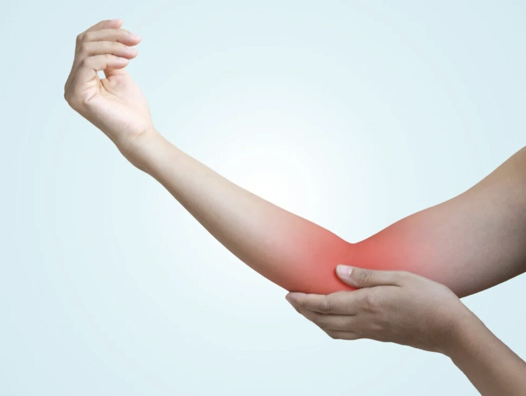
A sudden tearing sensation in the front of your elbow during a heavy lift often signals a distal biceps tendon rupture. This tendon connects the biceps muscle in your upper arm to the radius bone in your forearm at the elbow. When the tendon tears, it typically causes immediate pain, swelling, and a noticeable deformity where the biceps muscle retracts toward the shoulder, sometimes described as the “Popeye” sign.
Without surgical repair, significant reductions in overall arm strength and supination strength (the ability to rotate your palm upward) may occur. Surgery can restore the tendon to the bone using specialised fixation techniques, helping to preserve strength and function.
Risk Factors and Causes
Distal biceps tendon ruptures occur when excessive force is placed on the tendon beyond its capacity. The injury typically happens when you attempt to lift a heavy object with your arm in flexion while the elbow is forcefully extended. This mechanism creates a sudden eccentric contraction that exceeds the tendon’s tensile strength.
Common scenarios include:
- Lifting heavy objects at work
- Weightlifting exercises (particularly bicep curls with heavy weights)
- Catching falling objects
- Contact sports injuries
Predisposing factors:
- Male gender
- Age between 40 and 60 years
- Smoking (reduces tendon vascularity)
- Anabolic steroid use
- Previous corticosteroid injections near the tendon
- Dominant arm involvement
Diagnosis
Clinical examination provides the primary diagnosis. Your orthopaedic surgeon will assess for the classic triad: pain at the antecubital fossa (front of the elbow), weakness in elbow flexion and forearm supination, and a palpable gap where the tendon has retracted.
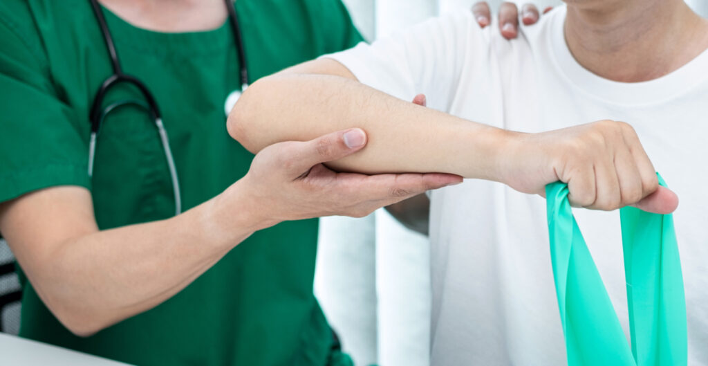
Physical examination tests:
- Hook test: The most reliable clinical test. Your surgeon attempts to hook their index finger under the biceps tendon from the lateral side with your elbow flexed at 90 degrees and your forearm supinated. An absent tendon indicates complete rupture.
- Biceps squeeze test: Squeezing the biceps muscle should produce elbow flexion if the tendon is intact.
- Passive forearm pronation test: Increased pronation compared to the uninjured side suggests rupture.
Imaging studies:
MRI confirms the diagnosis and evaluates retraction distance. Ultrasound serves as an alternative dynamic imaging modality, particularly useful for partial tears. X-rays rule out associated fractures but do not visualise soft tissue injuries.
Surgical Techniques
Surgery is performed under regional anaesthesia (nerve block) combined with general anaesthesia or sedation. The procedure typically takes 60-90 minutes.
Single-Incision Anterior Approach
Procedure:
A 4-5 cm incision is made in the antecubital fossa (front of the elbow). The surgeon identifies the retracted tendon, removes degenerative tissue, and prepares the footprint on the radial tuberosity. The lateral antebrachial cutaneous nerve and radial nerve are carefully protected.
Fixation methods:
- Cortical button technique: Sutures are passed through the tendon and connected to a button that flips on the opposite cortex of the radius
- Suture anchor fixation: Anchors are placed directly into the radial tuberosity, with sutures passed through the tendon
- Interference screw: The tendon is secured in a bone tunnel with a bioabsorbable or metal screw
Advantages:
Direct visualisation, shorter operative time, and a single surgical wound.
Risks:
Higher risk of heterotopic ossification (abnormal bone formation), potential radial nerve injury, and difficulty with chronic retractions.
Two-Incision Approach (Boyd-Anderson)
Procedure:
A smaller anterior incision identifies and retrieves the tendon. A second lateral incision over the radius allows direct visualisation of the radial tuberosity while the forearm is pronated, protecting the posterior interosseous nerve. The tendon is passed through a bone tunnel and secured.
Advantages:
Lower heterotopic ossification rates, enhanced protection of neurovascular structures, and better exposure for anatomic footprint restoration.
Disadvantages:
Longer operative time, two surgical incisions, and greater technical complexity.
Postoperative Care and Recovery
Weeks 0-2: Protection Phase
Immediate postoperative period:
- Your arm is immobilised in a hinged elbow brace locked at 90 degrees
- The forearm is positioned in neutral rotation
- Wrist and hand exercises begin immediately
- Ice therapy reduces swelling
- Pain management with prescribed analgesics
Goals:
Protect the repair, control inflammation, and prevent stiffness in adjacent joints.
Weeks 2-6: Early Mobilisation
Week 2:
- The elbow brace is unlocked to allow controlled passive range of motion
- Extension is limited to 30 degrees from full flexion initially
- Active elbow flexion begins with gravity eliminated (arm supported)
- Supination and pronation exercises start in mid-range
Week 4:
Extension progressed to neutral (0 degrees)
Active-assisted exercises introduced
Gentle isometric strengthening at mid-range positions
Week 6:
Full passive range of motion should be achieved
Active range of motion in all planes
Light resistance strengthening begins
Weeks 6-12: Strengthening Phase
Progressive loading:
- Resistance exercises gradually increase from 0.5 kg to 2-3 kg
- Emphasis on controlled eccentric strengthening
- Proprioception and coordination drills
- Gradual return to functional activities
Restrictions:
- No lifting objects heavier than 2-3 kg
- Avoid sudden jerking motions
- No contact sports or heavy labour
Months 3-6: Return to Function
Month 3:
- Full range of motion expected
- Strengthening progresses
- Sport-specific exercises begin for athletes
- Return to sedentary work
Month 4:
- Strengthening the contralateral side
- Return to light manual labour
- Progressive functional training
Month 6:
- Return to unrestricted activities
- Full strength is typically restored by 6-12 months
- Contact sports clearance with surgeon approval
Outcomes and Complications
Success rates:
Outcomes with restoration of strength and function when surgery is performed within 2-3 weeks of injury.
Potential complications:
Nerve injuries:
- Lateral antebrachial cutaneous nerve: Causes numbness over the lateral forearm
- Superficial radial nerve: Results in numbness over the dorsal thumb
- Posterior interosseous nerve: Motor loss is rare but can affect finger extension
Heterotopic ossification:
More common with the anterior single-incision approach. Usually asymptomatic, but may limit forearm rotation. Indomethacin prophylaxis or radiation therapy reduces risk.
Re-rupture:
Typically occurs from premature return to heavy activities or trauma during rehabilitation.
Stiffness:
Extension loss is more common than flexion limitations. Addressed with prolonged physiotherapy or manipulation under anaesthesia in resistant cases.
Synostosis:
Rare complication that eliminates forearm rotation. More common with the two-incision technique and heterotopic ossification.
Conservative Management vs Surgery
Non-operative treatment:
Reserved for low-demand elderly patients, significant medical comorbidities, or partial tears.
Functional deficits without surgery:
- Reduction in elbow flexion strength
- Reduction in supination strength
- Persistent deformity (Popeye sign)
- Fatigue with sustained forearm rotation activities
- Inability to perform heavy manual labour
Comparative outcomes:
Studies consistently demonstrate superior strength recovery, patient satisfaction, and return to pre-injury activity levels with surgical repair compared to non-operative management.
Return to Sports and Work
Sedentary work: 2-4 weeks
Light manual labour: 3-4 months
Heavy manual labour: 6 months
Non-contact sports:
- Swimming, cycling: 3 months
- Golf, tennis: 4-5 months
Contact sports:
- Full clearance: 6 months minimum
- Protected participation: Case-by-case evaluation
Strength testing criteria:
Return to unrestricted activity requires achieving strength greater than that of the contralateral side in flexion and supination, as measured by isokinetic dynamometry.
Conclusion
Distal biceps tendon rupture surgery restores tendon anatomy to the radius bone, preserving elbow flexion and forearm supination strength. The procedure involves either a single-incision or a two-incision surgical approach, with various fixation methods. Successful outcomes depend on surgical timing (within 2-3 weeks of injury) and adherence to structured rehabilitation protocols spanning 6 months for full recovery.
If you are experiencing sudden elbow pain following a lifting injury, visible biceps muscle deformity, or weakness in arm rotation and flexion, consult an orthopaedic surgeon immediately. Early surgical intervention within 2-3 weeks optimises outcomes and prevents permanent strength deficits.