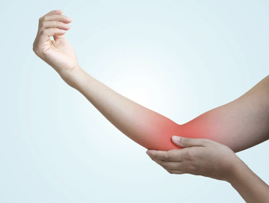
Did you know that the olecranon bursa can swell to the size of a golf ball within hours of injury or infection? The olecranon bursa (a small, fluid-filled cushion) sits directly over the bony tip of your elbow. When this fluid-filled sac becomes inflamed or infected, it swells noticeably, creating the characteristic “Popeye elbow” appearance.
Unlike joint swelling that restricts movement through internal pressure, olecranon bursitis typically allows full elbow motion. This happens because the bursa lies outside the joint capsule (the tissue that encloses the joint). The underlying cause—whether trauma, infection, or chronic irritation—determines the appropriate management approach. Recognising the difference between septic (infected) and aseptic (non-infected) bursitis early can help prevent complications and guide you towards the right care pathway.
Anatomy and Function of the Olecranon Bursa
The olecranon bursa normally contains a very small amount of synovial fluid (a lubricating liquid). This creates a friction-reducing layer that allows skin to glide smoothly over bone when you bend and straighten your elbow. This bursa develops after birth in response to mechanical stress. This explains why it’s particularly susceptible to irritation in adults who frequently lean on hard surfaces.
Bursal Structure
The bursa wall consists of synovial membrane cells (specialised cells that line the bursa). These cells can produce fluid rapidly when irritated. In healthy states, fluid production and absorption remain balanced. Inflammation disrupts this equilibrium. This leads to fluid accumulation that can expand the bursa significantly within hours of injury or infection.
Common Causes of Olecranon Bursitis
Repetitive Pressure and Trauma
Prolonged leaning on elbows creates microtrauma (minor repeated injuries) that triggers inflammation. This is frequently seen among students, office workers, and anyone who rests their elbows on desks. Single traumatic events like falling directly onto the elbow can cause immediate swelling with or without bleeding into the bursa.
Occupations requiring frequent crawling or kneeling with elbow contact carry a higher risk. Examples include plumbing or electrical work. Athletes in contact sports may develop bursitis from repeated falls or direct blows.
Infection (Septic Bursitis)
Bacteria can enter through small cuts, scrapes, or insect bites near the elbow and colonise the bursa. Staphylococcus aureus (a common bacterium) causes many cases of sepsis. The superficial location of the olecranon bursa—with minimal tissue protection—makes it vulnerable to penetrating injuries that introduce pathogens (disease-causing germs).
Septic bursitis progresses faster than aseptic forms. It requires different approaches to treatment for olecranon bursitis, typically involving antibiotics and sometimes a procedure to drain the fluid.
Underlying Medical Conditions
Gout (a condition in which uric acid crystals deposit in joints) and rheumatoid arthritis (an autoimmune disease that causes joint inflammation) can trigger bursitis. This happens through crystal deposition or autoimmune inflammation, respectively. These cases may present with symptoms throughout the body or involvement of other joints. This helps distinguish them from isolated mechanical causes.
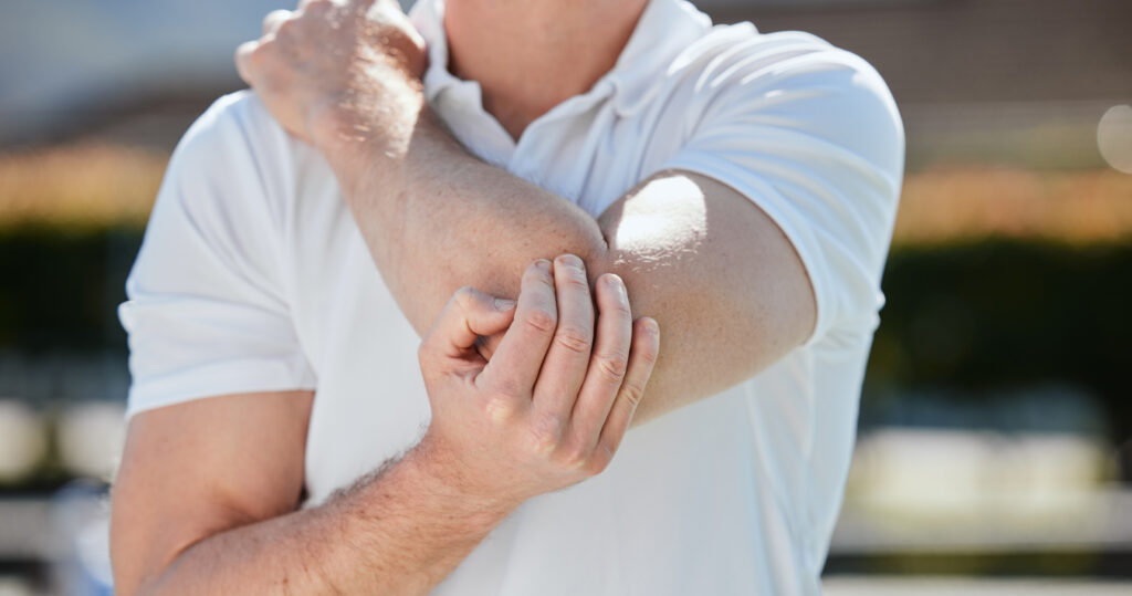
Recognising Symptoms and Warning Signs
Aseptic bursitis typically presents with visible swelling at the elbow tip, mild discomfort with direct pressure, and preserved range of motion. The swelling may fluctuate based on activity levels and usually feels soft or slightly firm.
Septic bursitis produces more concerning symptoms:
- Redness spreading beyond the immediate swelling
- Warmth is significantly greater than the opposite elbow
- Fever or feeling generally unwell
- Rapidly worsening pain
- Pain at rest rather than only with pressure
💡 Did You Know?
The olecranon bursa is one of many bursae in the human body, but its superficial position makes it among the more commonly infected bursae seen in clinical practice.
Diagnostic Approaches
Physical examination (where the doctor checks your elbow by looking at it and touching it) often provides sufficient information for initial management decisions. Comparing both elbows for temperature, redness distribution, and tenderness patterns helps distinguish septic from aseptic cases.
Aspiration and Fluid Analysis
When infection is suspected or the diagnosis remains unclear, needle aspiration can provide diagnostic clarity. This is a procedure where the doctor uses a needle to remove fluid from the bursa. Laboratory analysis examines:
- Cell count and type (the number and kind of cells in the fluid—elevated white cells suggest infection or inflammation)
- Gram stain and culture (tests that identify bacterial causes)
- Crystal analysis (looks for crystals that indicate gout or pseudogout)
- Glucose levels (sugar content in the fluid—reduced in infection)
Aspiration also serves therapeutic purposes. It immediately reduces swelling and pressure.
Imaging Studies
X-rays (images created using radiation to see inside the body) may reveal bone spurs, fractures, or foreign bodies that contributed to bursitis development. Ultrasound (imaging that uses sound waves) effectively visualises fluid collections and can guide aspiration procedures. MRI (magnetic resonance imaging—a detailed scan using magnets) is rarely necessary but helps evaluate complex cases or rule out other elbow pathology.
Conservative Olecranon Bursitis Treatment
Many aseptic cases respond well to non-surgical management over several weeks.
Activity Modification
Eliminating pressure on the affected elbow allows inflammation to subside. Using cushioned elbow pads when resting on surfaces, modifying work positions, and avoiding direct leaning helps prevent further irritation. Complete rest is unnecessary. Normal arm movement without direct pressure is encouraged.
Compression and Elevation
Elastic bandaging provides gentle compression that limits fluid accumulation. Keeping the elbow elevated above heart level when resting supports fluid drainage through gravity.
Ice Application
Applying ice wrapped in a cloth for intervals of fifteen to twenty minutes reduces inflammation and provides pain relief. Ice therapy is helpful during the acute phase (the early stage of swelling). It loses effectiveness for chronic cases.
Anti-Inflammatory Medications
Non-steroidal anti-inflammatory drugs (medications that reduce swelling and pain without steroids) reduce swelling and discomfort. Oral medications like ibuprofen or naproxen are commonly used for several days to weeks, depending on response. Topical anti-inflammatory gels offer an alternative with fewer systemic effects. The appropriate medication and dosage should be determined by a healthcare professional.
⚠️ Important Note
Never apply heat to a swollen elbow before infection has been ruled out—warmth accelerates bacterial growth and can worsen septic bursitis significantly.
Medical and Surgical Interventions
Aspiration Procedures
Removing accumulated fluid through needle aspiration provides immediate relief and allows fluid testing. The procedure takes only minutes. Your doctor inserts a needle to drain the swelling. This can be performed in clinic settings. Some physicians inject corticosteroids (anti-inflammatory medications) after aspiration to reduce inflammation. This approach carries a small risk of infection.
Fluid may reaccumulate after aspiration, sometimes requiring repeated procedures. Persistent recurrence despite conservative measures may indicate the need for further treatment.
Antibiotic Therapy
Septic bursitis requires antibiotic treatment. Treatment initially covers common pathogens until culture results guide specific therapy. Mild cases may respond to oral antibiotics. Severe infections need intravenous administration (medication given directly into a vein) and close monitoring.
Treatment duration typically extends over several weeks to ensure complete bacterial eradication. Inadequate antibiotic courses risk recurrence or chronic infection. A healthcare professional should determine the appropriate antibiotic regimen.
Surgical Bursectomy
When conservative treatment for olecranon bursitis fails after several months, or when infection cannot be controlled with antibiotics alone, surgical removal of the bursa may be necessary. Your surgeon can perform the procedure through an open incision (a cut in the skin) or arthroscopic techniques (using small instruments and a camera through tiny cuts). The approach depends on the clinical situation.
Post-operative recovery involves wound care, gradual return to activities, and elbow padding during healing. A new bursa typically regenerates over several months, usually without recurrent problems.
What Our Orthopaedic Specialists Observe
Delays in treating septic bursitis can lead to complications. These include abscess formation (a pocket of pus) or spread of infection to surrounding tissues.
Outcomes improve when patients consistently implement protective measures. This means not just during acute symptoms but as ongoing prevention. Workplace modifications and proper padding make the difference between single episodes and chronic recurring problems.
Putting This Into Practice
- Use elbow padding during activities: Gel or foam pads worn during desk work, exercise, or occupational tasks distribute pressure away from the bursa.
- Modify workstation ergonomics: Adjusting desk height, using armrests properly, and taking regular position changes reduces sustained elbow pressure.
- Treat skin injuries promptly: Cleaning and covering cuts or abrasions near the elbow helps prevent bacterial entry that could lead to septic bursitis.
- Manage underlying conditions: If gout or rheumatoid arthritis contributed to bursitis, work with your doctor to optimise treatment for these conditions. This may help reduce recurrence risk.
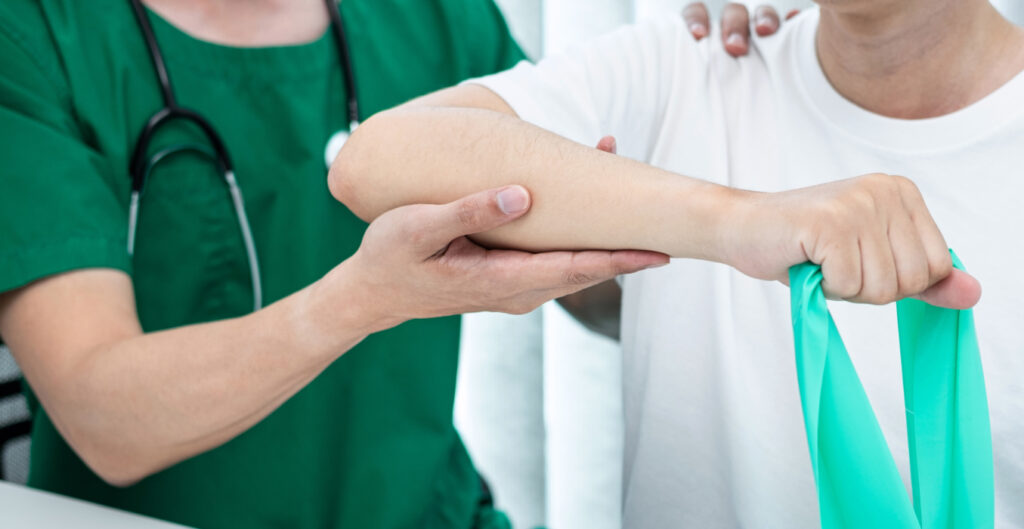
- Maintain flexibility: Gentle elbow stretching and strengthening exercises support joint health without stressing the bursa.
When to Seek Professional Help
- Swelling that persists beyond two weeks despite home care measures
- Redness, warmth, or spreading discolouration around the elbow
- Fever accompanying elbow swelling
- Inability to fully bend or straighten the elbow
- Pain that worsens progressively rather than improving
- Fluid that reaccumulates rapidly after draining
- Any open wound near a swollen bursa
Commonly Asked Questions
Can olecranon bursitis heal without treatment?
Mild aseptic bursitis often resolves with activity modification alone over several weeks. However, continuing activities that caused the initial inflammation may prevent healing. This may lead to chronic bursitis requiring medical intervention.
How do I know if my bursitis is infected?
Septic bursitis typically causes significant warmth, spreading redness, fever, and worsening pain even at rest. Aseptic bursitis usually causes swelling without these systemic signs. When uncertain, seek medical evaluation promptly. Delayed treatment of infection risks serious complications. Early detection allows for appropriate antibiotic treatment.
Will the swelling come back after aspiration?
Fluid commonly reaccumulates after aspiration. This happens because the underlying bursal inflammation persists. Addressing the root cause through activity modification and protective measures may help reduce the risk of recurrence. Multiple aspirations or surgery may be needed for persistent cases.
How long does recovery from bursectomy take?
Response times vary depending on your specific condition. Many patients return to normal activities within four to six weeks after the surgeon removes the bursa. Wound healing takes approximately two weeks. This is followed by gradual progression in activity. You should avoid heavy lifting or direct elbow pressure for six to eight weeks.
Can I exercise with olecranon bursitis?
Activities that don’t involve direct elbow pressure or impact are generally acceptable. Swimming, walking, and lower-body exercises typically cause no problems. Avoid push-ups, planks, or any exercise requiring elbow contact with surfaces until swelling resolves.
Next Steps
Aseptic bursitis typically resolves with activity modification and protective measures. Septic bursitis requires prompt antibiotic treatment to prevent complications. Persistent or recurrent swelling may need aspiration or surgical removal of the bursa.
If you’re experiencing persistent elbow swelling, redness, warmth, or pain that worsens at rest, consult an orthopaedic specialist to determine whether your condition requires medical intervention.