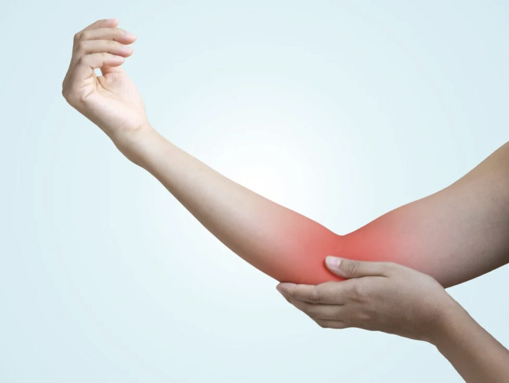
Did you know that most people who develop golfer’s elbow have never picked up a golf club? The condition develops when the tendons connecting your forearm muscles to the bony bump on the inside of your elbow become inflamed or develop small tears. Office workers, musicians, and tradespeople develop it as frequently as golfers. The pain typically starts at the medial epicondyle (the inner elbow) and can radiate down the forearm. This makes everyday activities like shaking hands, turning doorknobs, or lifting objects uncomfortable.
Anatomy of Medial Epicondylitis
The medial epicondyle serves as an anchor point for the flexor-pronator muscle group—five muscles responsible for bending your wrist, rotating your forearm palm-down, and gripping objects. These muscles share a common tendon attachment at this bony prominence. When you grip a golf club, twist a screwdriver, or type on a keyboard, these muscles contract and pull on this shared attachment point.
How Tendon Damage Occurs
Tendons consist of collagen fibres arranged in parallel bundles. They are designed to transmit force from muscle to bone. Repetitive stress causes microscopic tears faster than the body can repair them. Over time, this leads to tendinosis, a degenerative process where healthy collagen (the main structural protein in tendons) breaks down and becomes disorganised. Blood vessel and nerve growth into the damaged area contributes to ongoing pain, even at rest.
Many cases affect the dominant arm. This reflects the cumulative stress from repetitive activities performed primarily with one hand.
Common Causes and Risk Factors
Golfer’s elbow results from cumulative microtrauma rather than a single injury. Activities requiring repeated wrist flexion (bending your wrist forward) or forearm rotation place high demand on the medial epicondyle attachment.
Occupational activities frequently responsible include:
- Using hand tools (hammers, screwdrivers, pliers)
- Typing and mouse work for extended periods
- Assembly line work requiring repetitive gripping
- Cooking and food preparation
- Construction and carpentry
Sports-related causes extend beyond golf:
- Throwing sports (baseball, javelin)
- Racquet sports with improper technique
- Weight training, particularly pulling exercises
- Climbing and bouldering
Individual risk factors include age in middle adulthood, when the tendon blood supply naturally decreases. This makes repair less efficient. Inadequate warm-up, sudden increases in activity intensity, and poor technique amplify stress on the medial elbow structures.
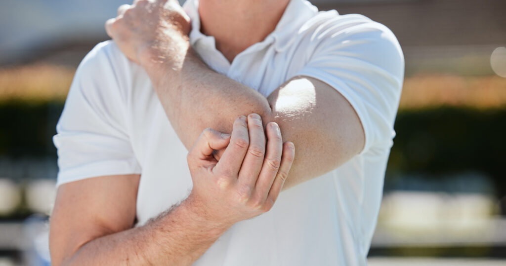
Recognising the Symptoms
Pain at the inner elbow that worsens with gripping is the hallmark symptom. This discomfort typically develops gradually over weeks to months rather than appearing suddenly.
Characteristic symptoms include:
- Tenderness directly over the medial epicondyle
- Pain when bending the wrist against resistance
- Discomfort when shaking hands or squeezing objects (such as opening jars, carrying bags, or using tools)
- Stiffness in the elbow, particularly in the morning
- Weakness in grip strength
- Numbness or tingling in the ring and little fingers (when the ulnar nerve is involved)
Symptoms often worsen with activity and improve with rest initially. As the condition progresses, pain may persist even during rest or sleep. The involvement of the ulnar nerve, which runs through a groove behind the medial epicondyle, can cause radiating symptoms into the hand.
Diagnostic Process
Clinical examination remains the primary diagnostic tool for identifying this condition (confirming whether you have golfer’s elbow based on your symptoms and physical findings). Your doctor will press on the medial epicondyle to reproduce tenderness and test resisted wrist flexion. Bending the wrist against resistance with the elbow straight typically reproduces or worsens the pain.
Physical Examination Tests
The resisted wrist flexion test involves fully extending your elbow whilst someone applies downward pressure as you try to flex your wrist. Pain at the medial epicondyle may indicate involvement of the flexor-pronator attachment.
Grip strength testing with a dynamometer (a device that measures how strongly you can squeeze) provides an objective measure and helps track treatment progress. Comparison with the unaffected arm establishes a baseline.
Imaging Studies
X-rays (images of bone structure) may be ordered to rule out other conditions, such as arthritis or loose bodies within the joint. They cannot visualise tendon damage directly.
Ultrasound (an imaging modality that uses sound waves to create images of soft tissues) allows real-time visualisation of tendon thickness, tears, and changes in blood flow. It can also guide injection therapies precisely to the affected area.
MRI (magnetic resonance imaging) creates detailed images of soft tissue structures using magnetic fields. It helps identify the extent of tendon damage, partial tears, or concurrent conditions affecting the elbow.
Non-Surgical Treatment Approaches
Many patients with golfer’s elbow respond to conservative treatment (non-surgical approaches such as rest, exercises, and lifestyle changes). Recovery typically requires several months. Tendon healing occurs slowly due to limited blood supply.
Activity Modification and Rest
Identifying and temporarily avoiding aggravating activities allows the healing process to begin. Complete immobilisation is unnecessary and may be counterproductive. Controlled movement maintains blood flow and prevents stiffness. Modifying technique, adjusting workstation ergonomics, or using different tools can reduce strain on the medial elbow.
Bracing and Support
A counterforce brace worn just below the elbow redirects forces away from the damaged tendon attachment. This reduces strain during activities and can allow continued function whilst healing progresses. Wrist splints worn at night prevent unconscious gripping during sleep.
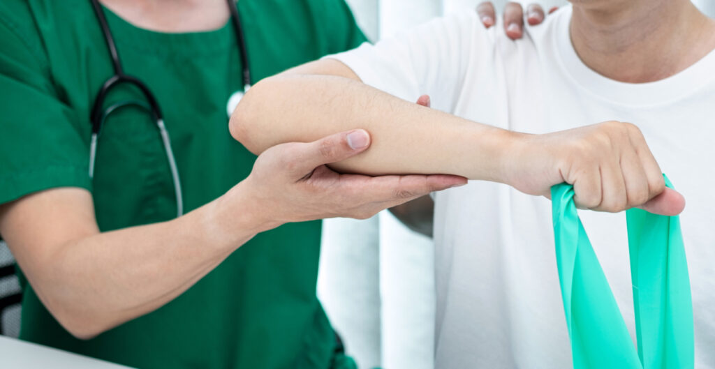
Physical Therapy
Structured rehabilitation forms an essential component of golfer’s elbow treatment. Therapy progresses through distinct phases:
- Pain management phase: Your physiotherapist uses ice, gentle stretching, and soft-tissue massage to reduce inflammation (swelling and irritation) in the affected area and restore flexibility.
- Strengthening phase: Your physiotherapist introduces eccentric exercises, slowly lowering a weight whilst the wrist extends. Eccentric loading (applying controlled stress as the muscle lengthens) stimulates collagen remodelling and tendon repair. This phase requires consistent effort over several weeks to produce structural changes.
- Functional rehabilitation: Your physiotherapist incorporates sport-specific or work-specific movements, gradually increasing load and complexity to prepare for a full return to activity.
💡 Did You Know?
Eccentric exercises work by applying controlled stress to healing tendons, stimulating the production of organised collagen fibres. This explains why complete rest often fails—tendons need appropriate loading to heal properly.
Medication
Oral anti-inflammatory medications (such as ibuprofen) provide short-term pain relief but do not accelerate healing. Topical anti-inflammatory gels applied directly over the medial epicondyle deliver medication locally with fewer systemic effects.
Short courses of medication can facilitate participation in physical therapy by managing pain during exercises.
Injection Therapies
When conservative measures provide insufficient relief, injection therapies offer additional options.
Corticosteroid Injections
Corticosteroid injections (medicines that reduce inflammation, delivered directly into the affected area) reduce inflammation and provide rapid pain relief, often within days. However, benefits typically last only a short period. Repeated injections may weaken tendon tissue. These injections are most useful as a bridge to facilitate physical therapy participation rather than as definitive treatment.
Platelet-Rich Plasma (PRP)
PRP therapy involves drawing a small amount of blood, concentrating the platelets (blood cells that contain growth factors), and injecting this preparation into the damaged tendon. Platelets contain growth factors that may enhance tissue healing. Some patients report sustained improvement following PRP treatment.
Prolotherapy
Prolotherapy uses dextrose (sugar) solutions to stimulate an inflammatory healing response. The mild irritation triggers the body’s repair mechanisms. This potentially strengthens weakened tendon attachments over multiple treatment sessions.
Surgical Treatment
Surgery becomes an option when symptoms persist despite several months of comprehensive non-surgical treatment. The procedure involves removing damaged tissue from the tendon attachment. In some cases, the surgeon releases a portion of the tendon to reduce tension.
What Surgery Involves
Your surgeon can operate through a small incision over the medial epicondyle or using arthroscopic techniques (a minimally invasive approach using a small camera and instruments inserted through tiny cuts). Your surgeon excises damaged, degenerative tissue. The remaining healthy tendon is reattached or allowed to heal in place. If ulnar nerve irritation accompanies golfer’s elbow, the surgeon may move the nerve to a more protected position.
Recovery After Surgery
Post-operative rehabilitation follows a structured timeline:
- Initial immobilisation in a splint for the first week or two
- Gradual range of motion exercises beginning around two weeks
- Strengthening exercises start around six weeks
- Return to full activity typically within several months
Compliance with rehabilitation protocols significantly influences surgical outcomes.
Prevention Strategies
Preventing recurrence requires addressing the factors that led to the initial injury.
Technique modification applies to sports and work activities. In golf, a proper grip, wrist position, and swing mechanics reduce medial elbow stress. In the workplace, ergonomic assessments can identify problematic movements or postures.
Gradual increases in activity intensity allow tendons to adapt. Sudden increases in training volume or work demands overwhelm the tissue’s ability to repair and strengthen.
Forearm conditioning through regular stretching and strengthening maintains tendon health. Eccentric exercises performed preventatively may reduce injury risk.
Appropriate equipment matters—using tools with the proper grip size, maintaining tennis or golf equipment, and ensuring an ergonomic workplace setup—contribute to reducing strain.
When to Seek Professional Help
- Pain persists for more than two weeks despite rest and home treatment
- Weakness in grip affects daily activities or work
- Numbness or tingling develops in the ring and little fingers
- Pain occurs at rest or disturbs sleep
- Symptoms worsen despite self-treatment efforts
- Elbow stiffness limits movement
Commonly Asked Questions
How long does golfer’s elbow take to heal?
With consistent conservative treatment, many cases improve within several months, though the timeline and degree of improvement vary from person to person. Mild cases may resolve within weeks. Severe or chronic cases can take longer. Tendon healing is inherently slow due to limited blood supply.
Can I continue exercising with golfer’s elbow?
You can maintain fitness through activities that don’t stress the medial elbow. Lower-body exercises, cardiovascular training, and upper-body movements that avoid gripping or wrist flexion are generally safe. Your physiotherapist can recommend specific modifications based on your activities.
What’s the difference between golfer’s elbow and tennis elbow?
Golfer’s elbow affects the inner (medial) elbow, where the wrist flexor tendons attach. Tennis elbow affects the outer (lateral) elbow, where the wrist extensor tendons attach. The conditions involve different muscle groups and result from different movement patterns. Treatment principles are similar.
Do I need an MRI to diagnose golfer’s elbow?
Clinical examination is usually sufficient for diagnosis. Your doctor may recommend an MRI or an ultrasound if symptoms are atypical, if other conditions need to be excluded, or if surgery is being considered. Imaging helps assess the extent of tendon damage and guides treatment decisions.
Will golfer’s elbow come back after treatment?
Recurrence is possible if you don’t address the underlying causes. Continuing preventive exercises, maintaining proper technique, and avoiding sudden increases in activity intensity help reduce the risk of recurrence. Some people remain more susceptible and may benefit from ongoing conditioning programmes.
⚠️ Important Note: Individual recovery experiences will differ due to personal health factors. This content is educational in nature and should not replace consultation with qualified healthcare professionals for tailored advice regarding your specific condition and treatment needs.
Next Steps
Conservative treatment resolves most cases when applied consistently. Structured physical therapy, activity modification, and gradual return to complete functional form are the foundation of recovery. Persistent symptoms beyond several months of appropriate treatment warrant professional evaluation.
If you’re experiencing persistent inner elbow pain, grip weakness, or numbness in your fingers, consult with an orthopaedic specialist to evaluate your condition and discuss treatment options.