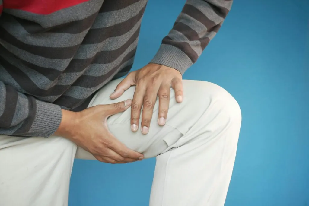
A Baker’s cyst, also called a popliteal cyst, is a fluid-filled swelling that develops behind the knee when synovial fluid accumulates in the popliteal bursa, a small sac that sits between the gastrocnemius and semimembranosus muscles. It forms through a one-way valve mechanism: fluid is pushed into the bursa during knee bending but cannot easily return to the joint, causing a gradual buildup that often worsens with activity. Importantly, the cyst is a symptom of an underlying knee condition, meaning treatment focused solely on the cyst, without addressing the root cause, frequently leads to recurrence.
How Baker’s Cysts Develop
Baker’s cysts form when excess synovial fluid, produced in response to joint irritation or inflammation, overflows into the popliteal bursa through a small opening in the posterior joint capsule. Common underlying causes include:
- Osteoarthritis: Cartilage breakdown triggers chronic low-grade inflammation, leading to increased fluid production that gradually fills the bursa.
- Meniscal Tears: Tears in the knee’s cartilage cushions create mechanical irritation and direct pathways for fluid to move into the popliteal bursa.
- Inflammatory Arthritis: Conditions such as rheumatoid arthritis, gout, and psoriatic arthritis generate significant synovial fluid during flares, accelerating cyst growth.
In younger patients without arthritis, a Baker’s cyst may signal underlying issues such as ligament injuries, cartilage damage, or other knee pathology warranting further investigation.
Recognising Baker’s Cyst Symptoms
The cyst typically presents as a soft, fluid-filled mass in the popliteal fossa, the diamond-shaped space behind the knee. Size varies considerably, from barely palpable to several centimetres in diameter. Smaller cysts may cause no symptoms whatsoever. Your doctor may discover them incidentally during imaging for other reasons.
As the cyst enlarges, patients commonly report:
- A sensation of fullness or tightness behind the knee, particularly noticeable when fully straightening or bending the leg
- Aching discomfort that worsens with prolonged standing or walking
- Visible swelling that may change size throughout the day or week
- Stiffness when initiating movement after periods of rest
The cyst’s relationship to the surrounding structures determines symptom severity. A moderate-sized cyst positioned against the tibial nerve (the major nerve running through the back of the knee) may cause more symptoms than a larger cyst sitting away from neurovascular structures. Compression of the popliteal vein can cause calf swelling mimicking deep vein thrombosis (a blood clot in a deep vein)—a clinical scenario that requires careful differentiation.
Pain directly from the cyst itself is relatively uncommon unless rupture occurs. Most discomfort stems from the underlying knee pathology generating the fluid. This explains why patients often experience anterior or medial knee pain alongside their posterior swelling.
Cyst Rupture: A Distinct Clinical Presentation
When a Baker’s cyst ruptures, synovial fluid dissects into the calf muscle compartments. This creates a characteristic clinical picture sometimes called “pseudothrombophlebitis.” The presentation can closely mimic deep vein thrombosis, so imaging is typically required to differentiate between them.
Ruptured cyst symptoms develop relatively quickly—often over hours—and include:
- Sudden sharp pain in the calf, sometimes described as a “pop” sensation
- Rapid calf swelling extending towards the ankle
- Ecchymosis (bruising) around the medial ankle, known as the “crescent sign”
- Difficulty bearing weight due to calf discomfort
Whilst rupture sounds alarming, it generally resolves with conservative management (non-surgical treatment). The body gradually reabsorbs the leaked fluid. However, distinguishing rupture from DVT remains clinically important given the vastly different treatment implications. Anticoagulation (blood-thinning medication) for DVT could worsen bleeding if the diagnosis is actually a ruptured cyst.
Diagnostic Approach
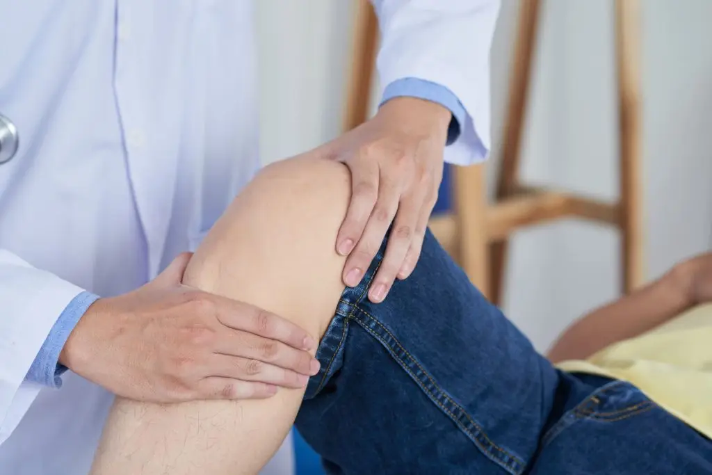
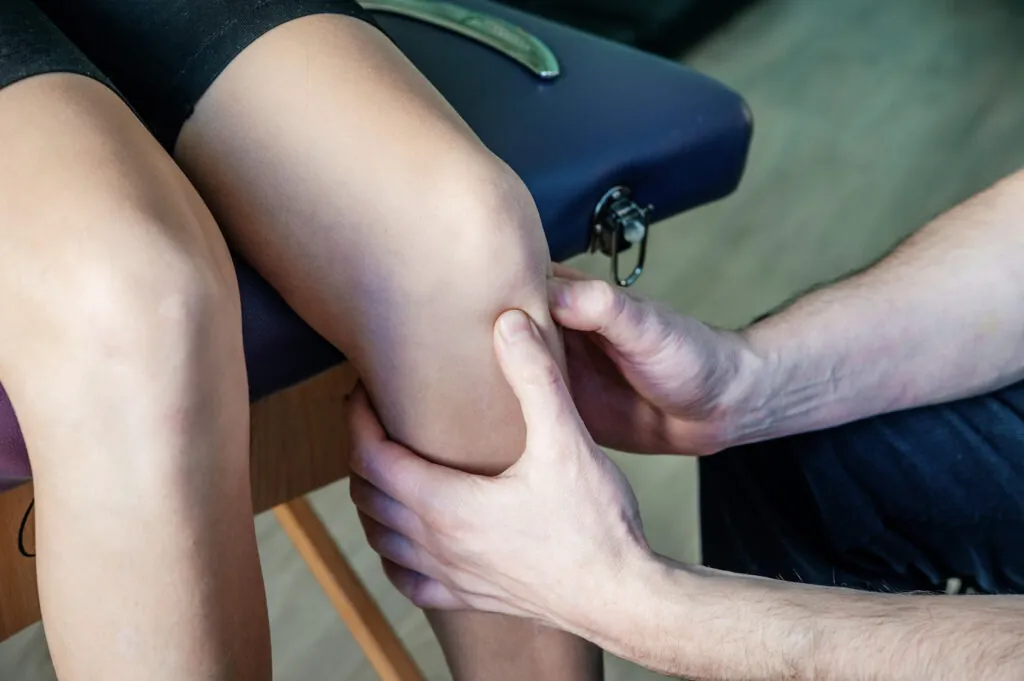
Clinical examination can provide substantial diagnostic information. The Foucher’s sign—a cyst that becomes firm and prominent with knee extension but soft and less distinct with flexion—helps confirm the diagnosis. It also helps differentiate the cyst from other popliteal masses.
Physical examination should assess:
- The cyst’s size, consistency, and exact location
- Range of motion limitations
- Signs of underlying knee pathology (effusion, ligament stability, meniscal signs)
- Calf examination for signs of rupture or venous compression
Ultrasound (a screening test that uses sound waves to create images of internal structures) offers real-time, non-invasive visualisation of the cyst. It confirms the cyst’s fluid-filled nature and demonstrates its communication with the joint space. Doppler ultrasound simultaneously evaluates the popliteal vessels, ruling out DVT when clinical suspicion exists.
MRI (Magnetic Resonance Imaging—a diagnostic test that creates detailed images using magnetic fields) provides a comprehensive evaluation when surgical planning is anticipated or when the underlying knee pathology requires detailed assessment. MRI reveals not only the cyst’s dimensions and relationships but also meniscal tears, cartilage lesions, and synovial abnormalities contributing to fluid production. For Baker’s cyst removal in Singapore, MRI findings guide surgical approach and help predict recurrence risk based on underlying pathology.
Non-Surgical Management
Conservative treatment aims to relieve symptoms and address the underlying cause of fluid production. Without addressing the source, the cyst typically refills even after draining. Your doctor will tailor the treatment approach based on your specific symptoms, activity level, and underlying knee condition.
Activity Modification
Reducing activities that stress the knee joint—particularly deep squatting, prolonged kneeling, and high-impact exercise—decreases synovial fluid production. This doesn’t mean immobilisation. Gentle movement and cycling are often well-tolerated whilst allowing the joint to settle.
Compression and Elevation
Elastic knee sleeves provide gentle compression that may limit cyst expansion. Elevating the leg above heart level when resting promotes fluid drainage from the knee and calf.
Physical Therapy
Strengthening the quadriceps and hamstrings improves joint stability. This potentially reduces mechanical irritation that drives fluid production. Therapy also addresses any gait abnormalities and compensates for knee dysfunction.
Aspiration
Needle aspiration (a procedure where your doctor inserts a needle to draw out fluid) removes fluid from the cyst. This provides temporary relief and confirms the diagnosis through fluid analysis. Aspiration alone has high recurrence rates because it doesn’t address the underlying cause or seal the communication with the joint.
Corticosteroid Injection
Your doctor may inject corticosteroid (an anti-inflammatory medication) into the cyst or joint space after aspiration. This reduces inflammation and may slow fluid reaccumulation. Combined aspiration and injection provides longer-lasting relief than aspiration alone. However, recurrence remains common if the underlying pathology persists.
Surgical Treatment Options
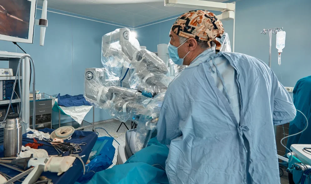
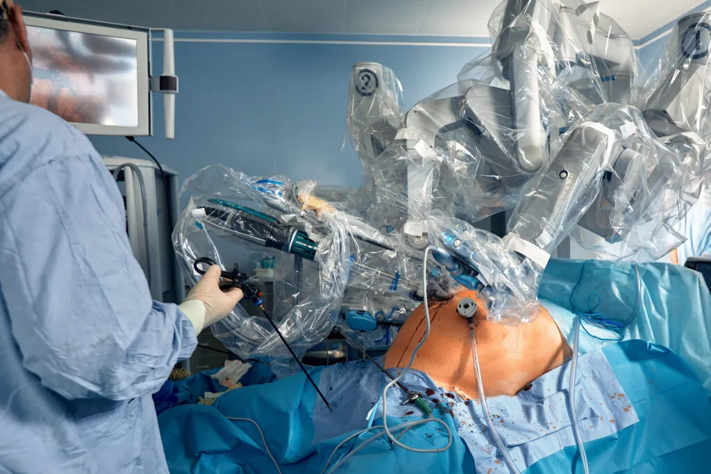
Surgery is considered when conservative measures fail, when the cyst causes significant functional symptoms, or when it compresses nearby nerves or blood vessels. Your orthopaedic surgeon will recommend the most appropriate approach based on your cyst size, underlying knee condition, and overall health.
Arthroscopic Treatment
Knee arthroscopy is a minimally invasive procedure that allows the surgeon to directly treat the source of excess fluid production — including meniscal repair, cartilage procedures, and synovectomy. Some surgeons also address the one-way valve mechanism arthroscopically, enlarging the communication between the joint and bursa to prevent fluid from accumulating.
Open Cyst Excision
Open cyst excision involves surgically removing the cyst through an incision at the back of the knee, typically recommended when the cyst is large, complex, or has not responded to previous treatments. Baker’s cyst removal in Singapore via open excision requires careful dissection, given the proximity to the popliteal vessels and tibial nerve.
Combined Approach
Many surgeons advocate combining arthroscopic treatment of underlying knee pathology with open or endoscopic cyst excision for more definitive management. This dual approach addresses both the root cause and the established cyst and is associated with lower recurrence rates than either method alone.
What Our Orthopaedic Surgeon Says
The clinical approach to Baker’s cysts has evolved over time. Rather than viewing the cyst as the primary problem requiring treatment, contemporary management recognises it as a symptom of underlying joint pathology. A thorough evaluation of the entire knee—not just the posterior swelling—guides treatment decisions.
Patients often present with a focus on the visible lump behind their knee. However, examination frequently reveals previously unrecognised joint-line tenderness, subtle effusion, or mechanical symptoms suggesting internal derangement. Addressing these findings typically provides more lasting relief than targeting the cyst directly.
Managing a Baker’s Cyst: Practical Steps
Monitor size changes by checking the popliteal area when your knee is extended. Compare to previous observations. Note whether symptoms correlate with activities or time of day.
Address modifiable factors contributing to knee inflammation. Maintain appropriate body weight to reduce joint stress. Choose lower-impact exercise options. Ensure proper footwear for your activities.
Manage flares conservatively with rest, ice application for 15-20 minutes several times daily, and over-the-counter anti-inflammatory medication if not contraindicated for you.
Pursue evaluation of underlying knee pathology rather than focusing solely on the cyst itself. Understanding why your knee produces excess fluid guides effective long-term management.
Communicate symptom changes to your healthcare provider, particularly new weakness, numbness, or sudden calf swelling that could indicate complications requiring prompt assessment.
When to Seek Professional Help
- Sudden severe calf pain with rapid swelling developing over hours
- Numbness, tingling, or weakness in the foot or lower leg
- A popliteal mass that feels firm, pulsatile, or is growing rapidly
- Calf swelling with skin warmth or redness, suggesting possible DVT
- Persistent symptoms despite several months of conservative management
- Knee pain or mechanical symptoms (such as catching, locking, or the knee giving way) alongside the cyst
- Any mass behind the knee in someone with a history of cancer
Commonly Asked Questions
Will my Baker’s cyst go away on its own?
Small cysts may resolve spontaneously, particularly if the underlying cause—such as a minor injury—heals. However, cysts associated with chronic conditions like osteoarthritis typically persist or fluctuate in size without treatment. The cyst follows the knee: when joint inflammation settles, the cyst often shrinks; when inflammation increases, the cyst enlarges.
Can I exercise with a Baker’s cyst?
Low-impact activities such as swimming, cycling, and walking are generally well-tolerated. Avoid deep squats, lunges, and high-impact activities that stress the knee joint and may increase fluid production. If exercise consistently worsens your symptoms, discuss activity modifications with your orthopaedic specialist.
How long is the recovery after Baker’s cyst surgery?
Recovery varies based on the procedure performed and whether concomitant knee pathology was addressed. Simple cyst excision may allow return to normal activities within several weeks. Combined procedures involving meniscal or cartilage surgery require longer rehabilitation—typically a few months before resuming sports. Your surgeon will provide specific recovery timelines based on your individual procedure.
Does draining the cyst cure it?
Aspiration provides temporary relief but doesn’t address the underlying cause of fluid production or the valve mechanism allowing fluid into the bursa. Recurrence rates after aspiration alone are high. Combined aspiration with corticosteroid injection offers somewhat longer-lasting results. However, definitive treatment usually requires addressing the underlying knee pathology.
Are there any risks if I leave the cyst untreated?
Small, asymptomatic cysts can often be monitored without intervention. Larger cysts carry risks of rupture (causing calf symptoms) and, rarely, compression of adjacent nerves or blood vessels. Leaving a cyst untreated doesn’t address the underlying knee condition, which may progress independently.
Next Steps
Baker’s cysts develop as a consequence of underlying knee pathology—treating the cyst alone without addressing the underlying cause results in recurrence. Aspiration combined with corticosteroid injection can provide interim relief, but definitive management typically requires identifying and treating the intra-articular condition driving excess fluid production. For persistent or enlarging cysts, combined arthroscopic and cyst excision procedures have lower recurrence rates than either approach alone.
If you are experiencing persistent swelling behind your knee, tightness or aching with knee movement, sudden calf pain, or associated mechanical symptoms such as catching or locking, consult an orthopaedic surgeon for evaluation and targeted treatment.