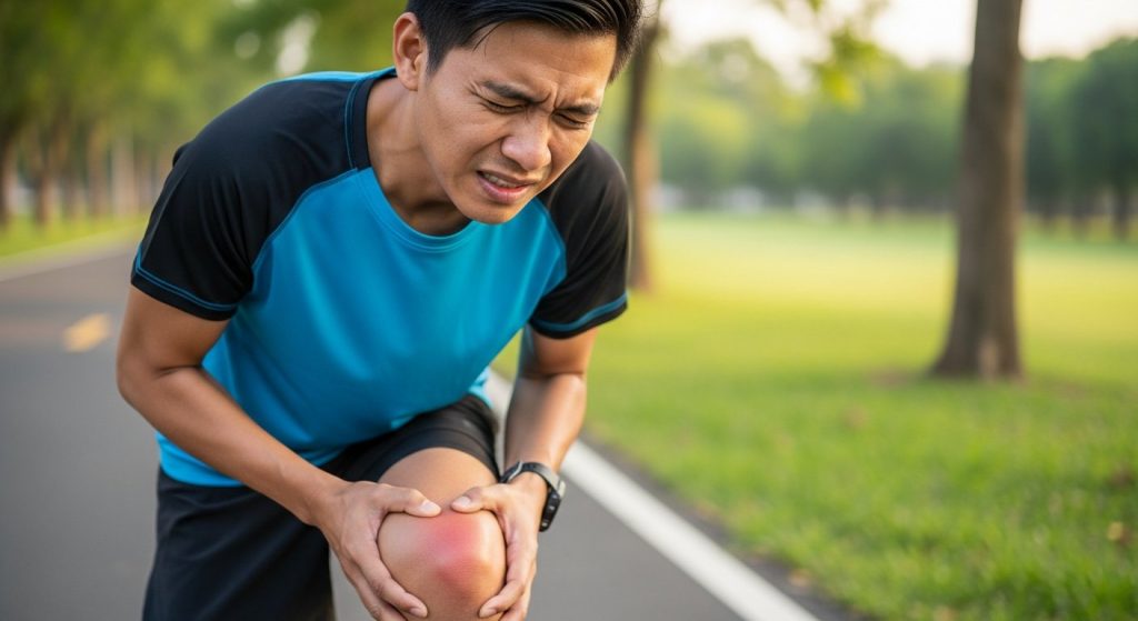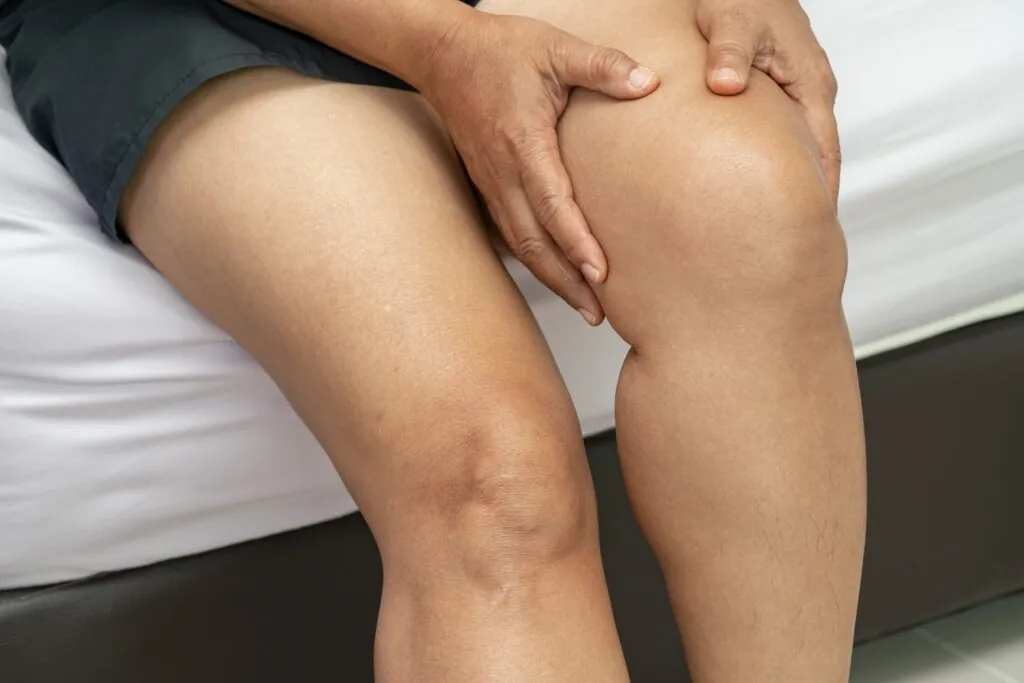
The kneecap, or patella, glides within a groove at the front of the femur during leg movement, and when it shifts partially or completely out of this groove, it tears the medial patellofemoral ligament in virtually every case, making this injury far more structurally significant than a simple sprain. This displacement most commonly occurs toward the outer side of the knee during twisting movements, sudden direction changes, or direct impact, causing immediate pain, swelling, and an inability to straighten the knee.
Patellar instability ranges from subluxation, where the kneecap partially shifts then returns, to complete dislocation requiring manual repositioning, with causes varying significantly between individuals. Understanding your specific anatomy and injury pattern determines which patellar dislocation treatment approach may offer reliable long-term stability.
Anatomy Contributing to Patellar Stability
Patellar stability depends on a combination of bony structure and soft tissue restraints working together to keep the kneecap tracking correctly through its groove. When any of these elements are compromised, whether through anatomy, injury, or both, the risk of displacement increases significantly.
- Trochlear groove: The patella sits within a channel formed by the lower femur; deeper grooves provide more bony constraint, while shallower grooves (trochlear dysplasia) offer less natural resistance to lateral displacement.
- Medial patellofemoral ligament (MPFL): This ligament connects the superomedial edge of the patella to the femur in the region between the adductor tubercle and medial femoral epicondyle.
- Quadriceps alignment and tibial tubercle position: The Q-angle indicates the direction of quadriceps pull, with higher angles creating greater lateral force on the patella; a laterally positioned tibial tubercle, where the patellar tendon attaches below the knee, further increases this stress during muscle contraction.
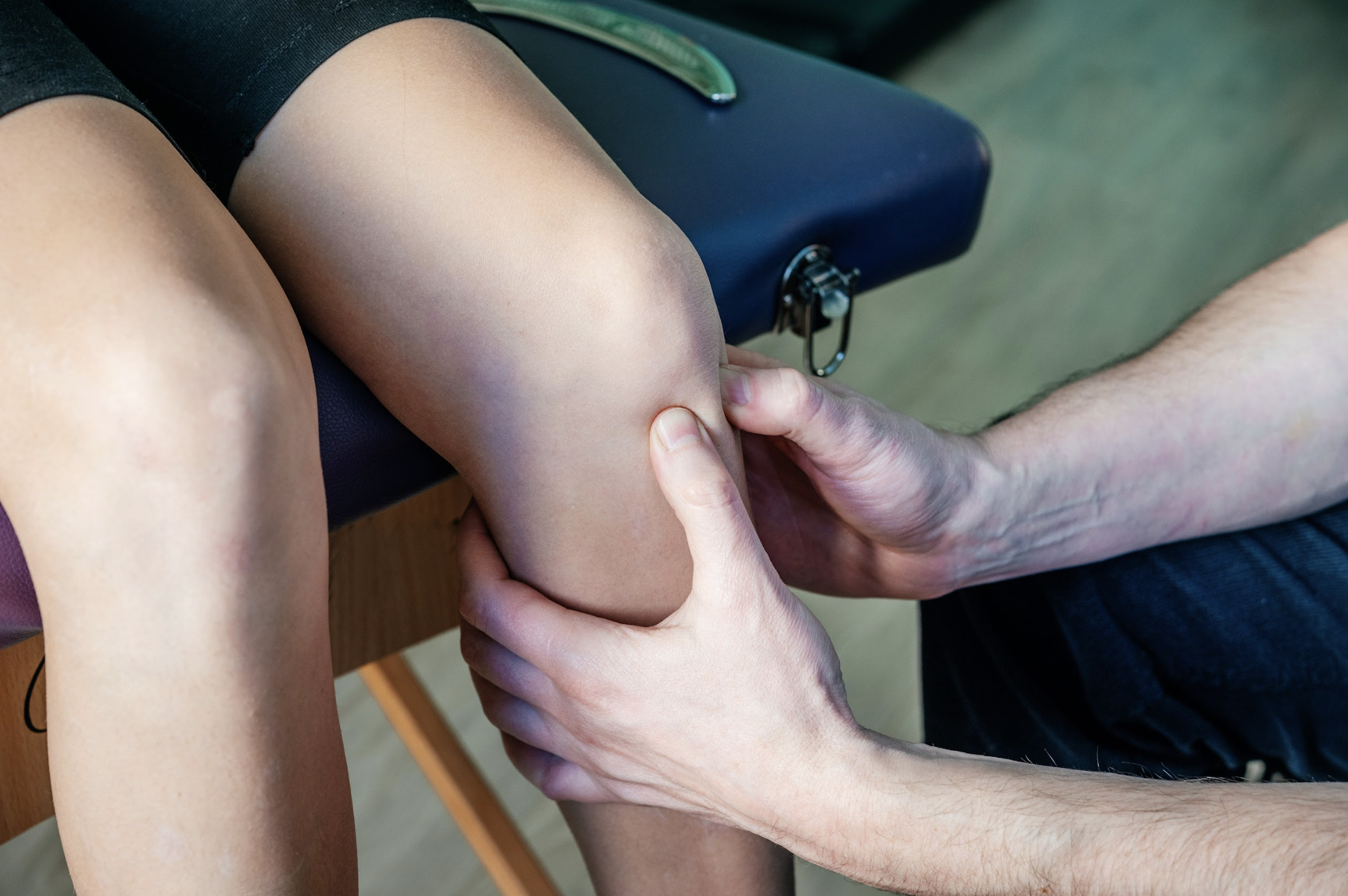
First-Time Dislocation Management
Initial management focuses on reducing swelling, restoring range of motion, and allowing soft tissue healing.
Your doctor may recommend a knee brace or immobiliser during the initial recovery phase to reduce pain and support the healing of the MPFL, though the type and duration of bracing varies by individual and clinical judgement, as current evidence does not clearly favour one bracing approach over another.
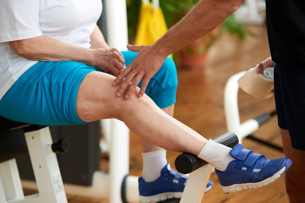
Physical therapy begins with quadriceps activation and straight-leg raises. It progresses to closed-chain strengthening as swelling resolves.
Quadriceps strengthening, including exercises targeting the vastus medialis obliquus (VMO), the inner quadriceps muscle, is commonly incorporated into rehabilitation, though its standalone role in preventing redislocation remains an area of ongoing clinical discussion.
Return to pivoting activities requires a full range of motion, quadriceps strength comparable to the uninjured side, and confidence with sport-specific movements. This timeline typically spans three to four months for non-operative management.
Risk Factors for Recurrent Instability
Several anatomical features increase the likelihood of repeated dislocations after initial injury. Identifying these factors helps determine which patients may benefit from early surgical consideration versus extended rehabilitation.
- Trochlear dysplasia describes a shallow or convex trochlear groove that provides inadequate bony constraint. Healthcare providers classify it from Type A (shallow but still concave trochlear groove) to Type D (severe dysplasia with a cliff-like step between facets).”
- Patella alta refers to a high-riding kneecap that sits above the trochlear groove during early knee flexion. This positioning means the patella lacks bony containment through the range where MPFL restraint is most critical.
- Tibial tubercle lateralisation positions the patellar tendon attachment point further toward the outer knee. This increases the lateral vector of quadriceps force. The tibial tubercle-trochlear groove (TT-TG) distance helps quantify this displacement. This is a measurement taken from imaging that shows how far the tendon attachment sits from the groove.
- Generalised ligamentous laxity affects overall joint restraint. This is where joints throughout the body are more flexible than typical. It may limit the effectiveness of a reconstructed or healed MPFL in stabilising the patella.
Age at first dislocation also influences outcomes. Younger patients, particularly those in early adolescence, demonstrate higher recurrence rates even with appropriate rehabilitation.
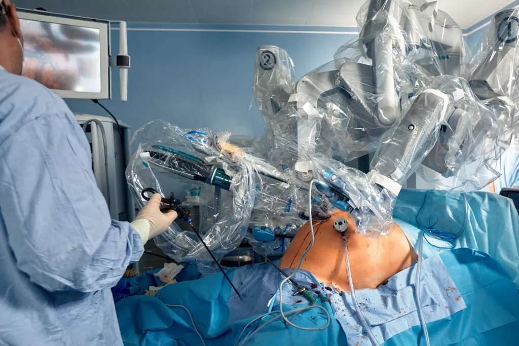
Surgical Treatment Options
When conservative management fails or anatomical risk factors suggest high recurrence likelihood, surgical intervention addresses the specific structural contributors to instability. Patellar dislocation treatment may combine multiple procedures tailored to your specific anatomy.
MPFL Reconstruction
The surgeon replaces the torn medial patellofemoral ligament with a tendon graft. The surgeon typically harvests it from the hamstrings or quadriceps. The graft is attached to the patella via bone tunnels or suture anchors. It fixes to the femur at the anatomic MPFL origin. Correct femoral tunnel positioning affects outcomes. Placement errors alter graft tension during knee motion and can alter patellofemoral contact pressure.
MPFL reconstruction has been shown to prevent redislocation in patients with relatively normal bony anatomy. When performed in isolation in patients with high-grade trochlear dysplasia, alongside other risk factors such as an elevated TT-TG distance or significant patella alta, recurrence rates are higher; however, isolated MPFL reconstruction may still be appropriate for patients with moderate dysplasia and no additional bony abnormalities.
Tibial Tubercle Osteotomy
During this procedure, the surgeon cuts and repositions the bone where the patellar tendon attaches. Moving the tubercle medially (toward midline) reduces the lateral vector of quadriceps pull. Anteriorisation shifts the tubercle forward, reducing patellofemoral contact pressures. Combined anteromedialisation addresses both vectors.
Tibial tubercle osteotomy suits patients with elevated TT-TG distance or those with patellofemoral articular cartilage damage, benefiting from pressure offloading. The bone cut requires screw fixation and creates some activity restrictions during healing.
Trochleoplasty
For severe trochlear dysplasia with a flat or convex groove, the surgeon deepens the trochlea to create bony containment where none existed. This procedure involves removing cartilage and bone, reshaping the groove, and repositioning the cartilage layer. In appropriately selected patients, outcomes show low recurrence rates.
Trochleoplasty is rarely performed in isolation; it is typically combined with MPFL reconstruction and, where indicated, tibial tubercle repositioning to address all contributing structural factors together.
Post-Surgical Rehabilitation
Recovery timelines depend on procedures performed and individual healing responses. MPFL reconstruction alone allows earlier motion than procedures involving bone cuts.
Weeks 1-6:
- Protected weight-bearing in a hinged brace locked in extension or limited flexion range
- Quadriceps activation and progressive range of motion within surgeon-specified limits; patellar mobilisation may be introduced at your surgeon’s discretion.
Weeks 6-12:
- Full weight-bearing, progressive strengthening, stationary cycling, and pool-based exercise
- Brace weaning typically occurs during this phase
Months 3-6:
- Sport-specific conditioning, agility progression, and confidence building
- Objective strength testing guides advancement decisions
Return to pivoting sports:
- Typically six to nine months post-surgery for MPFL reconstruction
- Potentially longer when osteotomy or trochleoplasty accompanies ligament reconstruction
Return to pivoting or competitive sports following MPFL reconstruction is typically guided by achievement of strength and functional milestones rather than a fixed timeframe. While some patients may progress toward sport-specific activities from nine months onwards, full return to play, particularly at a competitive level, may take considerably longer depending on individual progress and the demands of the sport.
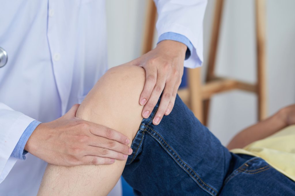
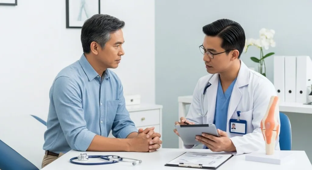
Preparing for Orthopaedic Consultation
Gathering relevant information before your appointment helps focus the evaluation on determining the appropriate treatment pathway.
Document your episodes:
- Note the circumstances of each dislocation or subluxation, including the activity, mechanism, whether the kneecap relocated spontaneously or required assistance, and recovery time after each event
Obtain prior imaging:
- Bring any X-rays or MRI scans from previous evaluations
- Comparison with new imaging helps assess progressive changes
List previous treatments:
- Include details of bracing, physical therapy duration and focus, and any activity modifications attempted
Note functional limitations:
- Describe which activities provoke symptoms, whether instability affects daily tasks or only sports, and your goals for treatment outcomes
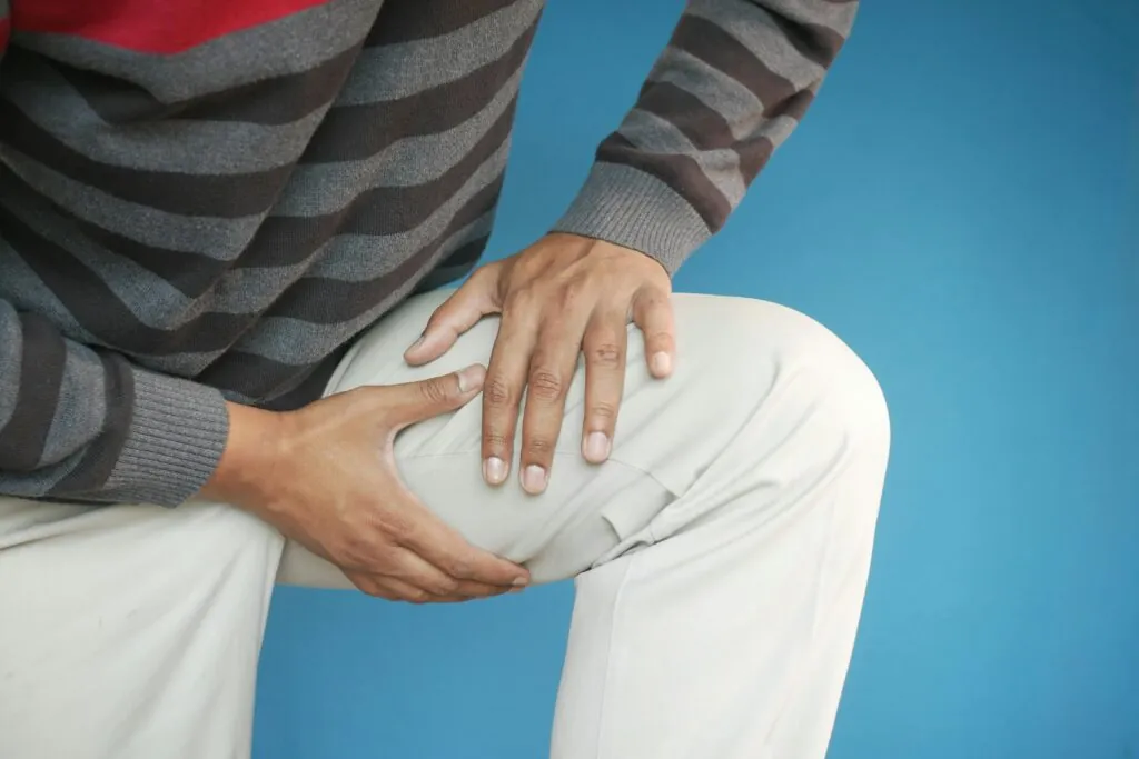
When to Seek Professional Help
- Knee gives way or feels unstable during walking or stair climbing
- Visible kneecap displacement that doesn’t immediately relocate
- Swelling persisting beyond two weeks after an instability episode
- Inability to fully straighten the knee after dislocation
- Multiple subluxation episodes despite rehabilitation
- Pain or grinding at the front of the knee with squatting or stairs
- Apprehension or avoidance of activities due to instability concerns
Commonly Asked Questions
Does a dislocated kneecap always require surgery?
First-time dislocations typically heal well with bracing and rehabilitation when no cartilage damage or loose fragments exist. Surgical consideration increases with recurrent episodes, anatomical risk factors suggesting high redislocation likelihood, or associated osteochondral injuries requiring repair.
How long until I can return to sports after patellar dislocation treatment?
Non-operative rehabilitation typically allows return to pivoting sports in three to four months. Surgical treatment extends this timeline to six to nine months. Exact duration depends on procedures performed and individual progress through rehabilitation milestones.
Will my kneecap dislocate again after surgery?
Outcomes differ among patients based on individual health factors and whether all contributing anatomical factors are addressed. Procedures that correct only part of the problem show higher failure rates.
Can I prevent patellar instability from developing?
When structural risk factors exist, prevention focuses on maintaining quadriceps and hip strength to optimise dynamic patellar control. Avoiding positions that place the patella at risk during pivoting activities may reduce but cannot eliminate dislocation risk when significant bony abnormalities exist.
Next Steps
First-time dislocations warrant thorough rehabilitation and imaging to identify structural risk factors, such as trochlear dysplasia, patella alta, or an elevated TT-TG distance. Recurrent instability despite appropriate conservative care requires surgical evaluation, as procedures must be matched to your specific anatomy; addressing only part of the problem substantially increases failure rates. Return to pivoting sports following surgery typically takes 6 to 9 months.
If you are experiencing recurrent kneecap dislocations, persistent knee instability, or apprehension during pivoting activities, consult with an orthopaedic surgeon for assessment and treatment tailored to your anatomy.