When a complete quadriceps tendon rupture occurs, the knee typically loses the ability to straighten against gravity, a functional deficit that generally requires surgical evaluation and intervention to restore it. The quadriceps tendon connects the four muscles at the front of your thigh to the kneecap, enabling leg extension for essential movements like walking, climbing stairs, and rising from a seated position. Quadriceps tendon repair involves surgically reattaching the torn tendon to the patella, followed by structured rehabilitation aiming to restore strength and movement.
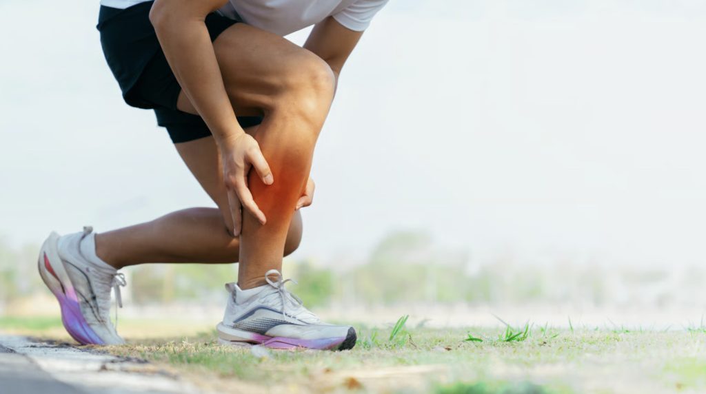
Many ruptures occur at the tendon-bone junction where the quadriceps meets the superior pole of the patella. The injury frequently results from a sudden eccentric load, such as landing from a jump or stumbling, where the quadriceps muscle contracts forcefully while the knee bends. Complete ruptures usually happen suddenly, often presenting with a noticeable pop followed by rapid swelling, localised pain, and a limited ability to perform a straight leg raise.
Recognising a Complete Rupture
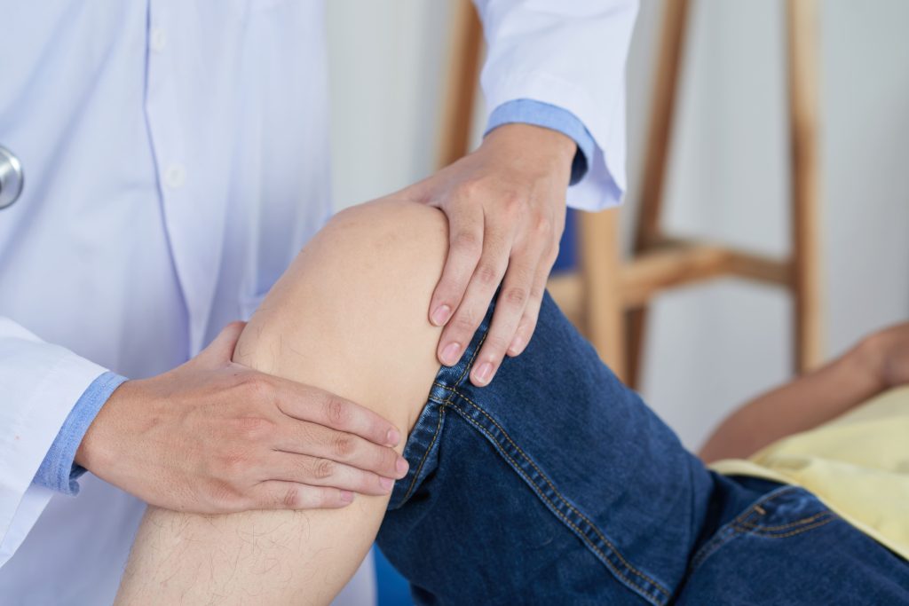
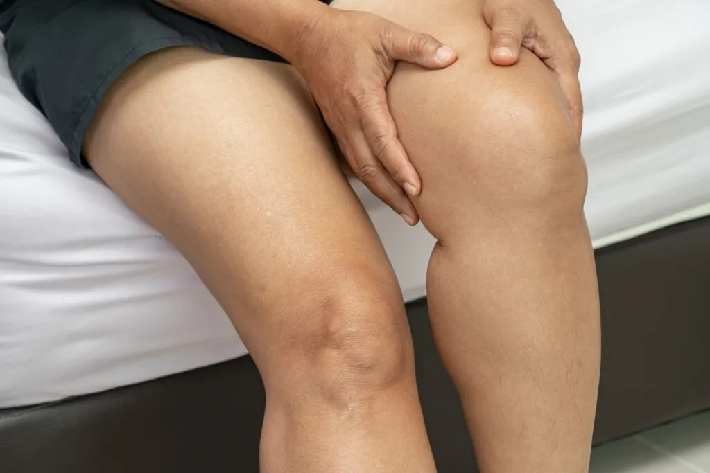
A complete quadriceps tendon rupture typically presents with distinctive clinical signs that help differentiate it from partial tears or other knee injuries. A common finding is a significant difficulty or inability to extend the knee against gravity, or to maintain knee extension when the leg is passively straightened. When lying flat and attempting to lift the leg while keeping the knee straight, the affected limb generally struggles to do so.
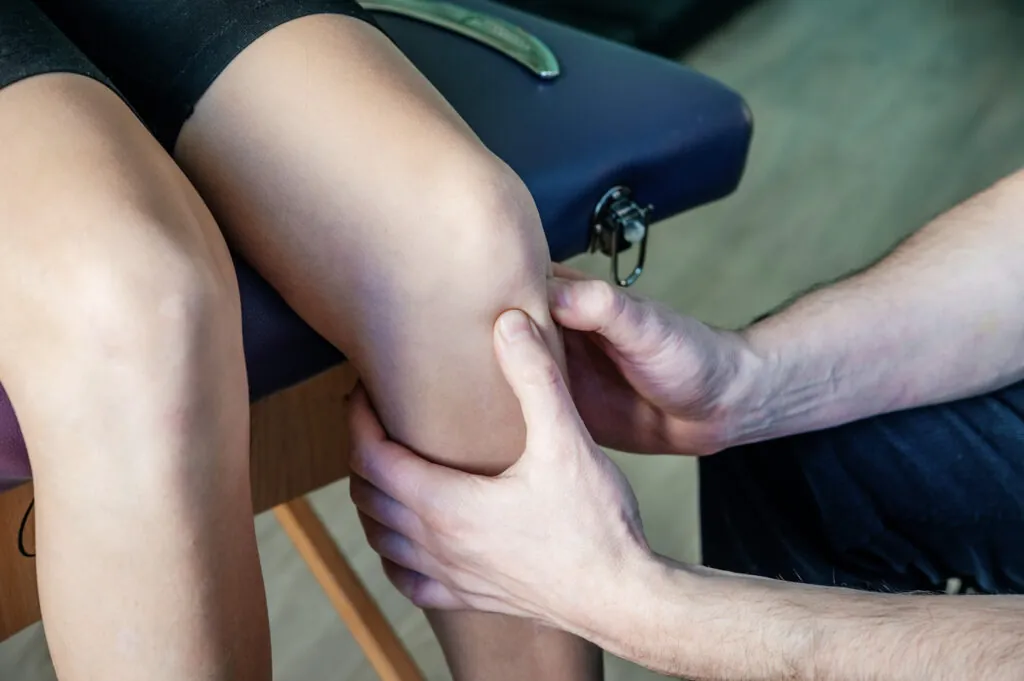
Physical examination often reveals a palpable gap or defect above the kneecap where the tendon normally resides. Swelling frequently develops within the first few hours, which can obscure this gap if the clinical examination is delayed. The patella may sit lower than normal (patella baja) because the quadriceps muscle tends to retract upward without its tendon anchor. Bruising typically appears within 24–48 hours, often spreading from the knee area down toward the shin.
Why Surgery Is Typically Necessary
Non-operative management is rarely the primary approach for complete quadriceps tendon ruptures because the torn ends tend to retract, hindering their ability to heal in a position that restores extension power. Without surgical repair, prolonged quadriceps weakness can significantly alter normal walking mechanics. The knee may buckle during the stance phase, navigating stairs can become highly challenging without handrail support, and rising from chairs frequently requires arm assistance.
Surgery aims to restore the anatomical attachment point and appropriate tension of the quadriceps mechanism. Clinical observations suggest that repairs performed within a few weeks of the injury generally achieve more predictable outcomes than delayed interventions. When surgery occurs months after the event, the tendon can retract significantly, and muscle atrophy may develop, making the surgical repair technically more demanding.
Surgical Repair Techniques
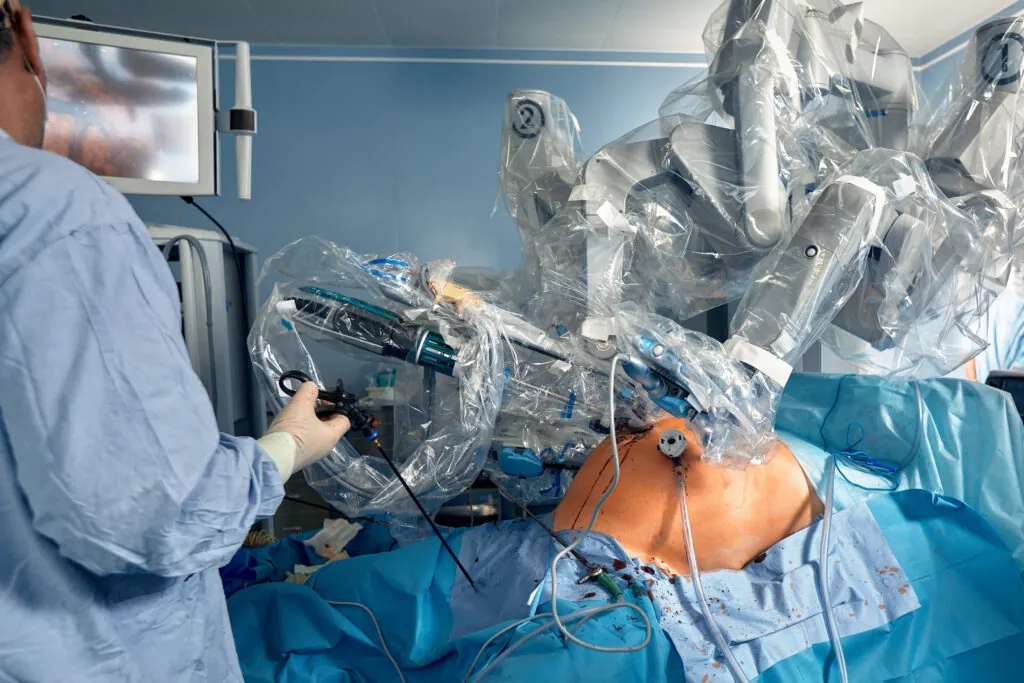
The standard approach typically involves making an incision over the front of the knee to expose the ruptured tendon and patella.
This differs from minimally invasive procedures such as standard knee arthroscopy, which is generally not utilised for repairing a complete, retracted tendon rupture. The surgeon identifies the torn tendon ends, manages any damaged tissue, and prepares the superior pole of the patella to create a suitable bone surface for tendon reattachment.
Suture anchor fixation involves utilising small anchors inserted into the patella with attached sutures. These sutures pass through the tendon in a structured pattern, often a locking configuration, designed to distribute tension across the repair site.
Transosseous tunnel repair involves creating small channels through the patella and passing sutures from the tendon through these tunnels to secure them. This technique is designed to provide stable fixation and remains a widely accepted option, particularly when baseline bone quality is favourable.
Immediate Post-Operative Care
Following surgery, the knee is immobilised in a hinged brace locked in full extension. This position protects the repair by minimising tension on the healing tendon. Weight-bearing begins immediately in most protocols. Patients walk with crutches whilst keeping the brace locked straight during the stance phase.
Wound care involves keeping the incision clean and dry until sutures or staples are removed, typically at 10-14 days. Applying ice and elevating the leg reduces swelling. Blood clot prevention measures include ankle pumps, calf squeezes, and sometimes medication, depending on individual risk factors.
Pain management combines scheduled anti-inflammatory medications with stronger analgesics for breakthrough pain. Most patients find pain manageable after the first week, with a gradual reduction in medication needs.
💡 Did You Know?
The intact extensor mechanism withstands substantial forces, several times body weight, during activities like stair descent, squatting, and rising from a chair. This is a pattern that surgical repair aims to restore, but that takes months of healing to approximate.
Rehabilitation Phases
Protection Phase
The knee remains braced in full extension except during supervised exercises. Quadriceps setting, isometric contractions whilst lying flat, begin immediately to maintain muscle activation without stressing the repair. Straight leg raises commence once quadriceps control allows lifting the leg without brace support, typically by day 3-5.
Passive range of motion exercises start under physiotherapy guidance. The therapist gently bends the knee within prescribed limits, usually 30-45 degrees initially, then gradually increases this range. Patella mobilisation prevents adhesions that could limit future movement.
Early Motion Phase
Brace settings progressively unlock to allow controlled flexion during walking. The typical progression increases permitted flexion by 15-20 degrees weekly, aiming for 90 degrees by week 6. Active-assisted knee bending exercises, using the opposite leg or a towel to help, complement passive motion.
Weight-bearing increases as quadriceps strength improves. Patients progress from two crutches to one, then to no assistive device, typically by weeks 4-6. The criterion for crutch weaning is adequate quadriceps control to prevent knee buckling during walking.
Strengthening Phase
Full range of motion becomes the goal, with particular attention to the final degrees of flexion. Closed-chain exercises, where the foot stays planted, include mini-squats, leg press, and step exercises. These movements strengthen the quadriceps whilst controlling patellofemoral joint forces.
Open-chain knee extension exercises (sitting leg raises with resistance) begin cautiously, often with a restricted range to protect the healing tendon. Progressive resistance follows individual tolerance and healing assessment.
Functional Restoration
Sport-specific or occupation-specific training begins for those returning to demanding physical activities. This includes agility drills, jumping progressions (starting with low amplitude), and endurance training. Running typically begins around months 4-5, starting with jogging intervals.
Strength testing compares the surgical leg to the opposite leg. Return to full activity generally requires quadriceps strength at 85-90% of the uninjured side, adequate range of motion, and confidence in functional movements.
Expected Recovery Timeline
Most patients return to desk work within 2-3 weeks, though mobility remains limited. Driving resumes once the brace allows adequate knee bending and the patient can perform emergency braking, typically 6-8 weeks for right leg injuries, sooner for left leg injuries in automatic vehicles.
Walking without a limp usually takes 8-12 weeks. Normal stair climbing, alternating feet without rail dependence, takes 3-4 months. Running and jumping activities require 4-6 months minimum, with full return to sport at 6-9 months.
⚠️ Important Note Attempting to progress faster than tissue healing permits risks repair failure. The tendon-bone junction takes approximately 12 weeks to achieve sufficient strength, regardless of how strong the repair feels subjectively.
Potential Complications
Re-rupture occurs in a small proportion of repairs, usually during the first 3 months when patients exceed activity restrictions or fall. Protecting the repair during the vulnerable healing period reduces this risk.
Stiffness develops when scar tissue limits knee bending. Patients who struggle to achieve 90 degrees of flexion by week 6 may require more intensive physiotherapy or, rarely, manipulation under anaesthesia. Consistent completion of prescribed exercises minimises this risk.
Quadriceps weakness persists to some degree in most patients long-term. Dedicated strengthening over 6-12 months maximises recovery, though subtle strength deficits may remain compared to the pre-injury state.
Infection, blood clots, and anaesthetic complications represent general surgical risks that appropriate perioperative care addresses.
Factors Affecting Outcomes
- Injury chronicity influences results. Acute repairs (within 2 weeks) generally achieve better functional scores and patient satisfaction than delayed repairs.
- Tendon quality at the time of surgery affects healing potential. Degenerative changes within the tendon, more common in older patients or those with prior tendinopathy, compromise tissue holding strength.
- Rehabilitation compliance determines functional outcomes more than surgical technique. Patients who complete prescribed exercises, attend physiotherapy sessions, and respect activity restrictions achieve better results than those who do not, regardless of repair method.
- Overall health influences healing capacity. Diabetes, smoking, chronic corticosteroid use, and kidney disease impair tissue repair and increase complication rates.
Putting This Into Practice
- Attend every physiotherapy session during the first 12 weeks when motion recovery is time-sensitive. Missed sessions create gaps in progression that become harder to correct later.
- Perform home exercises twice daily as prescribed. The in-clinic sessions establish technique, but home practice drives actual improvement.
- Monitor for warning signs, including increasing pain, sudden loss of motion, wound drainage, or giving way episodes. Report these promptly rather than waiting for the next scheduled appointment.
- Arrange your environment before surgery: place frequently used items at waist height, clear floor obstacles, secure loose rugs, and install temporary stair rails if needed.
- Plan work modifications realistically. Even desk workers need provisions for leg elevation and movement breaks during the early weeks.
When to Seek Professional Help
- Inability to straighten your knee or perform a straight leg raise after a knee injury
- A palpable gap or depression above your kneecap following sudden pain
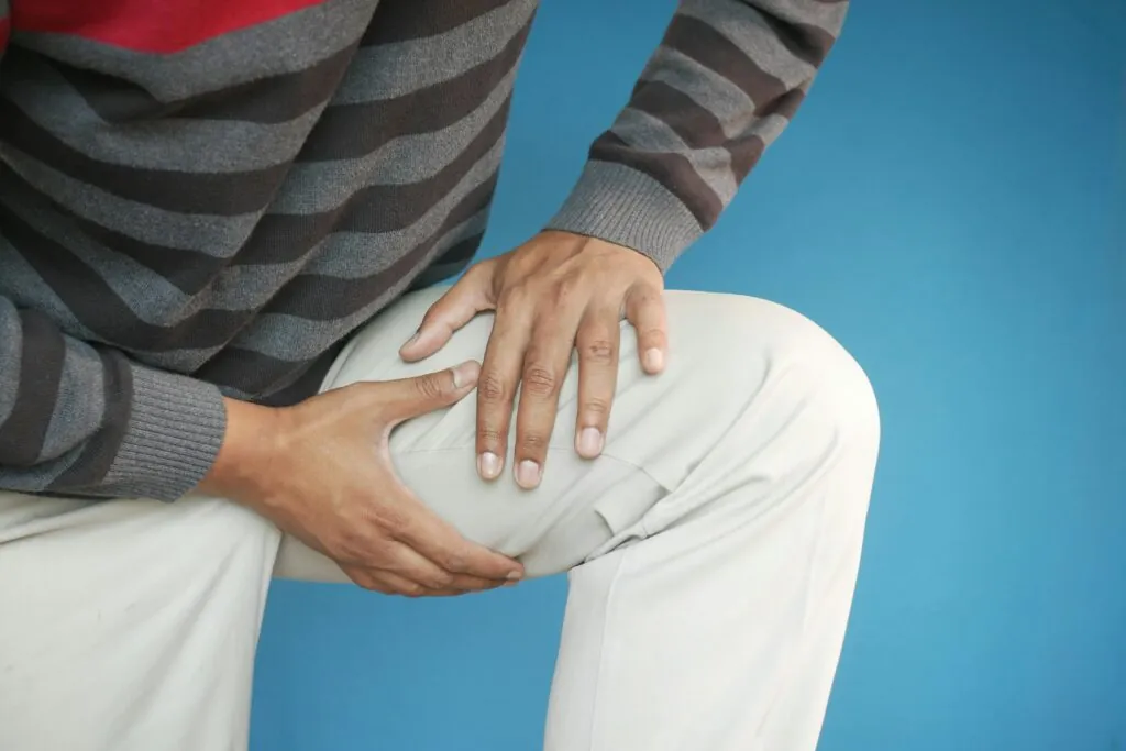
- Knee buckling or giving way during walking or climbing stairs
- Persistent swelling and weakness weeks after a suspected tendon injury
- Concern about re-injury or unusual symptoms during rehabilitation
Commonly Asked Questions
How long will I need to wear the knee brace?
Most protocols require the brace for 6-8 weeks, though restrictions gradually relax during this period. Initially locked in extension, the brace progressively unlocks to allow increased flexion as healing permits. The surgeon determines discontinuation timing based on clinical examination and healing assessment.
Can the tendon rupture again after repair?
Re-rupture occurs in a small proportion of repairs, most commonly within the first 3 months. Adhering to activity restrictions during early healing reduces this risk. Once fully healed and rehabilitated, the repaired tendon functions reliably for normal activities, though extreme loading scenarios carry some ongoing risk.
Will I be able to return to sports after surgery?
Most patients return to recreational sports between 6 and 9 months post-operatively. Return depends on achieving adequate strength, motion, and confidence rather than arbitrary timelines. High-impact activities like basketball or tennis require longer rehabilitation than cycling or swimming.
What happens if I delay surgery?
Delays beyond 2-3 weeks result in tendon retraction and early scar formation, making repair more difficult. Delays of several months often require tendon reconstruction with graft tissue rather than primary repair. Functional outcomes decline with increasing delay, making early surgical consultation advisable.
How much physiotherapy will I need?
Supervised physiotherapy typically involves 2-3 sessions weekly for the first 6 weeks, then 1-2 sessions weekly for another 6-8 weeks. Total duration extends 4-6 months for most patients, though those returning to demanding activities may continue longer. Home exercise compliance influences how much supervised therapy is needed.
Next Steps
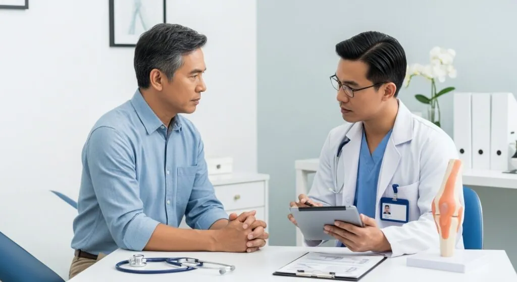
Clinical evidence indicates that addressing a complete rupture relatively early can support more predictable functional outcomes than a delayed repair. Long-term recovery depends heavily on rehabilitation compliance, which includes attending supervised physiotherapy sessions, completing prescribed home exercises, and respecting activity restrictions to protect the healing tendon-bone interface. Because this junction typically requires around 12 weeks to achieve sufficient baseline strength, progressing ahead of the recommended timeline can increase the risk of repair failure.
If you are experiencing an inability to straighten your knee, a palpable gap above your kneecap, or knee buckling following a sudden injury, consulting an orthopaedic surgeon in Singapore can provide a prompt evaluation to explore your treatment options.