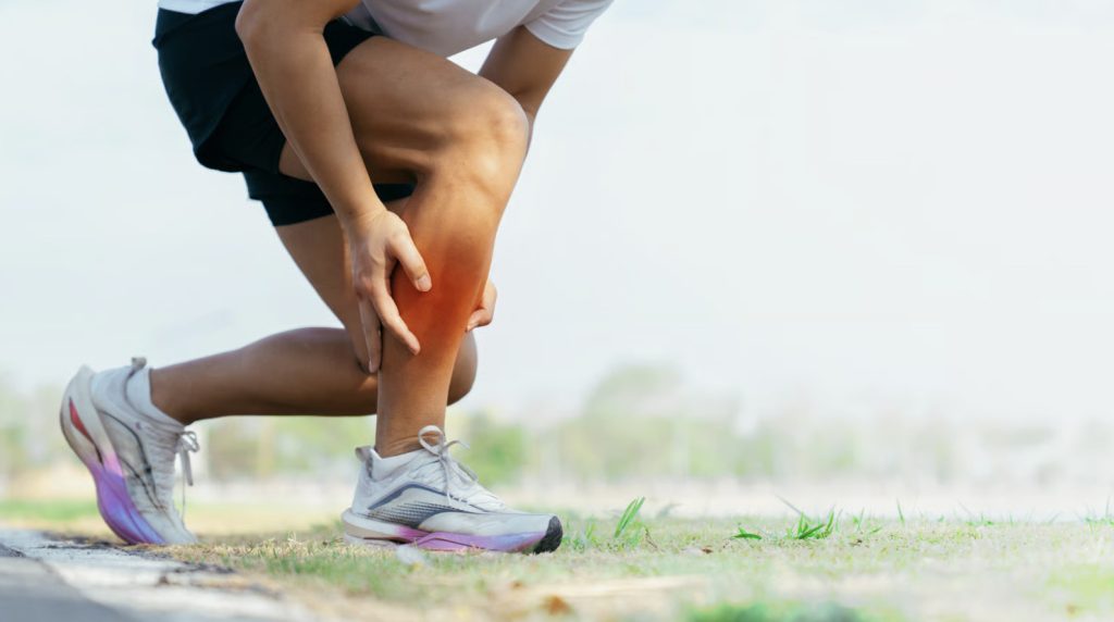
The infrapatellar fat pad contains one of the highest concentrations of nerve endings in the knee, making it exceptionally sensitive to the compression and irritation that occurs when it becomes pinched between the kneecap and thighbone. This structure, also known as Hoffa’s fat pad, sits beneath the kneecap and behind the patellar tendon, changing shape continuously during knee flexion and extension as it glides out of the way of moving joint surfaces.
When the fat pad becomes enlarged through inflammation or fails to move properly, repeated pinching occurs during routine movements like walking, climbing stairs, or straightening the leg fully, typically producing the pain and functional limitation characteristic of Hoffa’s fat pad impingement.
The condition often coexists with other knee pathologies, making accurate identification important for appropriate management.
Anatomy and Function of the Infrapatellar Fat Pad
The infrapatellar fat pad occupies the space between the patellar tendon anteriorly, the tibia inferiorly, and the femoral condyles posteriorly. This triangular structure fills what would otherwise be dead space within the knee joint, providing mechanical cushioning and contributing to joint lubrication through synovial fluid distribution.
Three distinct portions make up the fat pad: the anterior subcutaneous portion, the middle portion behind the patellar tendon, and the posterior portion that extends into the joint space. The posterior portion is most frequently involved in impingement syndromes because of its proximity to articular surfaces.
Blood supply to the fat pad comes from branches of the inferior genicular arteries, making it a highly vascularised structure that responds readily to injury with swelling and inflammation. This rich blood supply, combined with dense nerve innervation, may explain why fat pad pathology often produces such pronounced pain responses compared to other soft tissue knee injuries.
During normal knee function, the fat pad deforms and redistributes with each movement cycle. Full extension pushes the fat pad anteriorly and superiorly, while flexion allows it to settle posteriorly. Disruption of this normal movement pattern—whether from altered biomechanics, direct trauma, or chronic irritation—can initiate the impingement cycle.
Recognising Impingement Symptoms
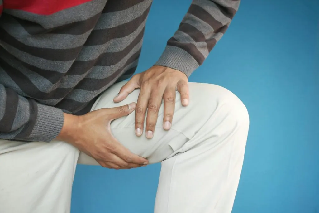
Anterior knee pain localised below and around the kneecap is often considered the primary symptom of Hoffa’s fat pad impingement. This pain typically worsens with activities requiring full knee extension, such as walking downhill, kicking, or holding the leg straight against resistance.
Patients frequently describe a sharp, pinching sensation at the front of the knee when moving from a bent to straight position. The pain often intensifies after prolonged sitting with knees bent, then standing—a pattern sometimes confused with patellofemoral syndrome but distinguished by its precise location inferior to the patella.
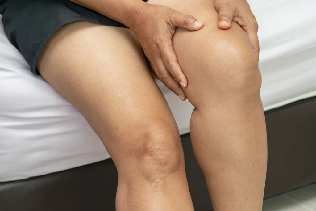
Swelling concentrated below the kneecap, visible as fullness on either side of the patellar tendon, suggests fat pad inflammation. This swelling differs from generalised knee effusion, which distributes throughout the joint space and obscures the normal contours around the kneecap.
Patients may notice their knee feels “blocked” or catches when approaching full extension, as though something is preventing complete straightening. This mechanical symptom typically results from the enlarged fat pad physically obstructing normal joint movement.
Pain Patterns and Aggravating Factors
Standing for extended periods typically aggravates symptoms because the fat pad remains compressed in the extended knee position. Running and jumping can produce intermittent sharp pains with each landing as the fat pad gets repeatedly pinched.
Hyperextension activities—common in dancers, gymnasts, and martial artists—place particular stress on Hoffa’s fat pad by maximising compression at end-range extension. Athletes in these disciplines may be more susceptible to developing impingement than those in sports requiring primarily flexed knee positions.
Night pain is uncommon with isolated fat pad impingement, helping differentiate it from inflammatory arthropathies or bone pathology. Pain that disturbs sleep warrants investigation for alternative or concurrent diagnoses.
Causes and Contributing Factors
Direct trauma to the front of the knee can initiate many cases of Hoffa’s fat pad impingement. Dashboard injuries, falls onto the knee, and direct blows during contact sports can cause bleeding within the fat pad, leading to scarring, fibrosis, and chronic enlargement that predisposes to impingement.
Repetitive microtrauma from overuse affects athletes and workers whose activities involve frequent full extension movements. The cumulative effect of thousands of minor pinching episodes can lead to gradual fat pad inflammation and hypertrophy.
Biomechanical factors play a substantial role in predisposing certain individuals to impingement. Genu recurvatum (knee hyperextension), quadriceps weakness allowing excessive anterior tibial translation, and patella alta (high-riding kneecap) all increase mechanical stress on the fat pad.
Post-Surgical Considerations
Knee surgery, particularly anterior cruciate ligament reconstruction and arthroscopic procedures, can trigger fat pad impingement through direct surgical trauma or altered joint mechanics during rehabilitation. The fat pad may become scarred to surrounding structures, limiting its normal mobility.
Aggressive extension exercises early in post-surgical rehabilitation sometimes precipitate fat pad problems. The inflamed, healing fat pad lacks its normal compliance and may become pinched during extension activities before adequate healing has occurred.
Surgical hardware positioning, especially tibial tunnel placement in ACL reconstruction, occasionally contributes to chronic fat pad irritation. Metal screws or fixation devices can impinge on fat pad tissue during specific movement ranges.
💡 Did You Know?
The infrapatellar fat pad can enlarge significantly when chronically inflamed, fundamentally changing its relationship with surrounding structures and potentially perpetuating the impingement cycle even after the original injury has healed.
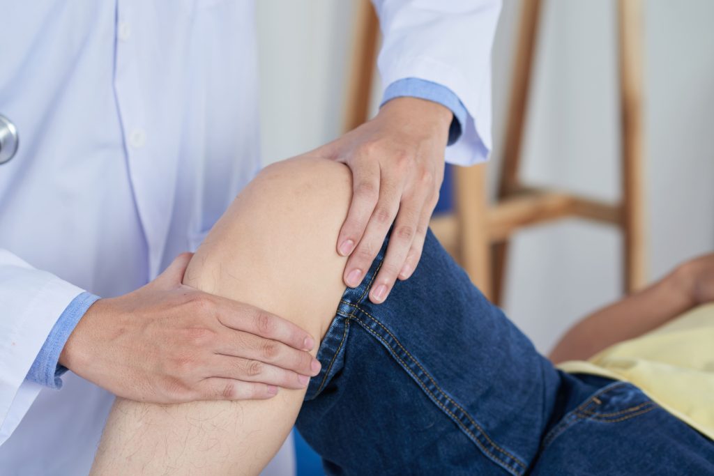
Clinical Assessment Methods
Physical examination begins with observation of standing posture and gait patterns. Hyperextended knees in standing and quadriceps avoidance patterns during walking suggest possible fat pad involvement.
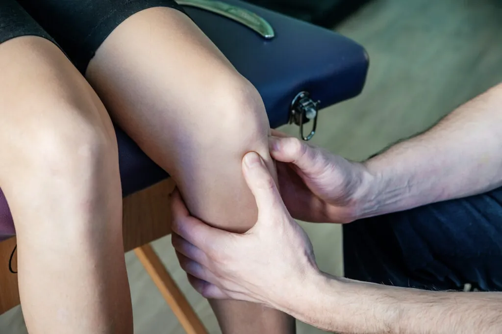
Palpation of the fat pad on either side of the patellar tendon with the knee slightly flexed often reveals localised tenderness in affected patients. Pressing into this soft tissue while the patient extends the knee may reproduce the catching pain characteristic of impingement.
The Hoffa test involves extending the knee while applying pressure to the fat pad. Reproduction of the patient’s typical pain suggests a positive test. Sensitivity improves when performed with the hip in neutral rotation, as internal or external hip rotation alters patellar tracking and may produce false results.
Range of motion assessment typically shows full flexion but painful or guarded terminal extension. Passive hyperextension testing reproduces symptoms and may be limited compared to the unaffected side.
Imaging Findings
MRI is generally considered a highly detailed method for fat pad visualisation, showing increased signal intensity on fluid-sensitive sequences when inflammation is present. Chronic cases demonstrate areas of fibrosis appearing as low signal bands within the normally homogeneous fat pad structure.
The characteristic “oedema pattern” on MRI shows high signal in the superoposterior aspect of the fat pad. This specific fluid distribution pattern can help support a diagnosis of impingement rather than simple contusion.
Lateral radiographs may show soft tissue density increase in the Hoffa’s fat pad region, though this finding is subtle and often missed. More importantly, X-rays evaluate for contributing factors like patella alta, osteophytes, or malalignment.
Ultrasound offers dynamic assessment, visualising fat pad movement during knee flexion and extension. Areas of scarring or abnormal motion can be identified in real-time, though operator experience significantly affects diagnostic accuracy.
Treatment Approaches
Initial management focuses on reducing inflammation and avoiding aggravating activities. Relative rest from extension-loading activities—rather than complete immobilisation—allows healing while maintaining joint mobility and muscle strength.
Ice application to the anterior knee for fifteen to twenty minutes several times daily helps control acute inflammation. Positioning the ice pack directly over the tender fat pad, easily accessible with the knee slightly bent, aims to optimise the therapeutic effect.
Non-steroidal anti-inflammatory medications may provide symptomatic relief and address the underlying inflammatory component. Short courses of oral NSAIDs or topical preparations applied directly to the anterior knee may help reduce pain and swelling during the acute phase. The appropriate medication, dosage, and duration should be determined by a healthcare professional.
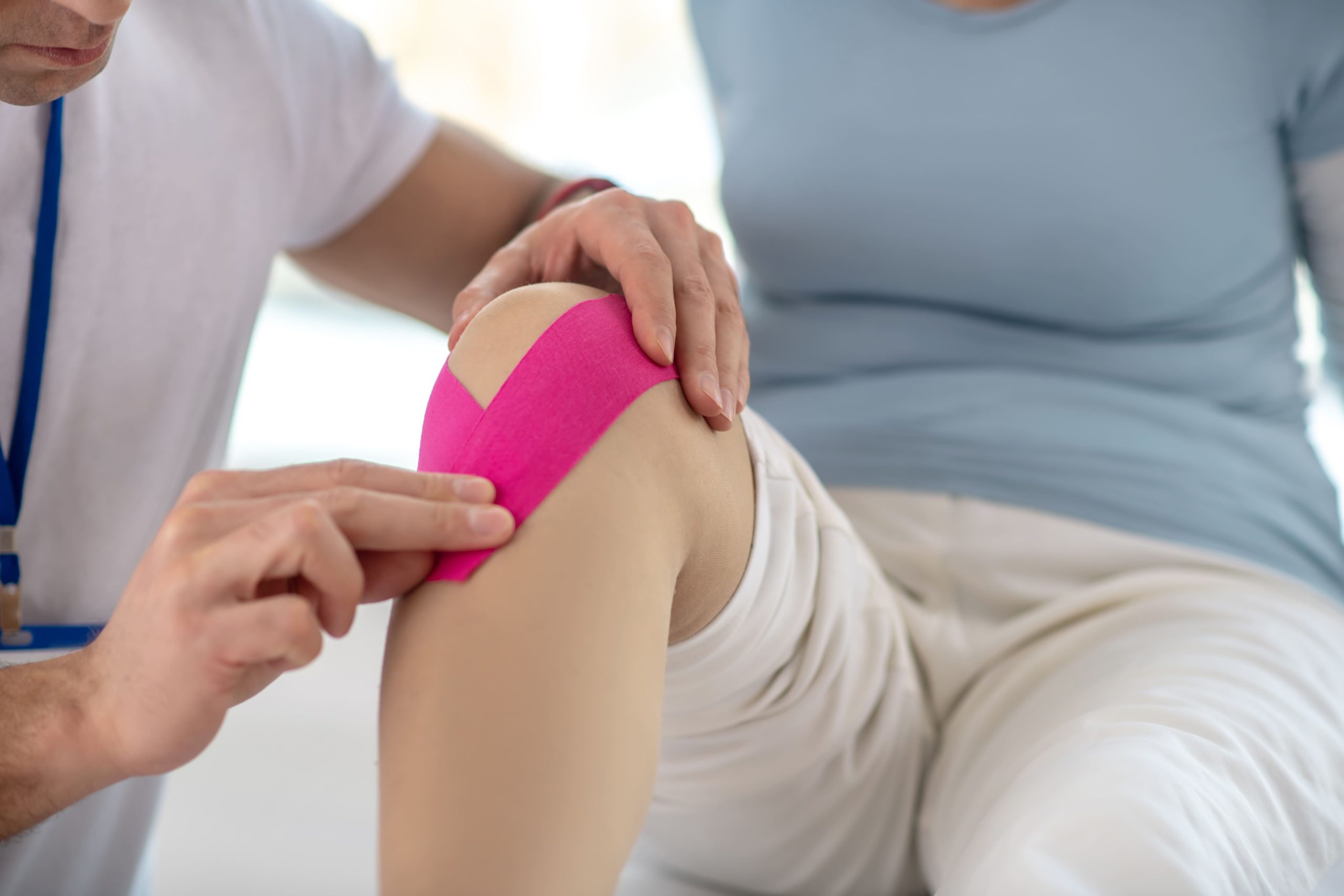
Taping techniques that lift the inferior pole of the patella may help reduce mechanical compression of the fat pad during activities. A simple strip of rigid tape applied from below the kneecap angling superiorly can provide symptom relief.
Physiotherapy Interventions
Quadriceps strengthening, particularly of the vastus medialis obliquus, can improve patellar tracking and may reduce fat pad stress. Exercises performed in partial flexion ranges initially avoid terminal extension until inflammation subsides.
Hip strengthening addresses proximal contributors to abnormal knee loading. Weakness in hip abductors and external rotators allows excessive femoral internal rotation, altering patellofemoral mechanics and increasing fat pad compression.
Manual therapy techniques including soft tissue mobilisation of the fat pad itself can restore normal tissue compliance. Gentle cross-friction massage aims to break down adhesions and improve fat pad mobility within the joint space.
Movement retraining addresses contributing biomechanical patterns. Athletes who hyperextend during landing or push-off require neuromuscular education to control knee position throughout functional activities.
⚠️ Important Note
Avoid aggressive stretching into knee hyperextension during rehabilitation—this may worsen fat pad compression and potentially delay healing. Range of motion exercises should stop at neutral extension until symptoms resolve.
Injection Therapies
Corticosteroid injection into the fat pad provides anti-inflammatory effects for cases not responding to conservative measures. The injection targets the fat pad directly, usually approaching from the inferolateral aspect of the knee to avoid the patellar tendon.
Pain relief following corticosteroid injection may begin within days and can last several months. This window may allow more effective rehabilitation, as patients may be able to perform strengthening exercises with reduced pain inhibition.
Repeated steroid injections carry risks of fat pad atrophy and weakening of adjacent soft tissues. Most practitioners limit injections to two or three over twelve months, reserving them for significant flare-ups rather than maintenance treatment.
Hyaluronic acid injections, while primarily used for osteoarthritis, may benefit some patients with Hoffa’s fat pad impingement by improving joint lubrication and reducing mechanical irritation. Evidence for this application remains limited compared to corticosteroids.
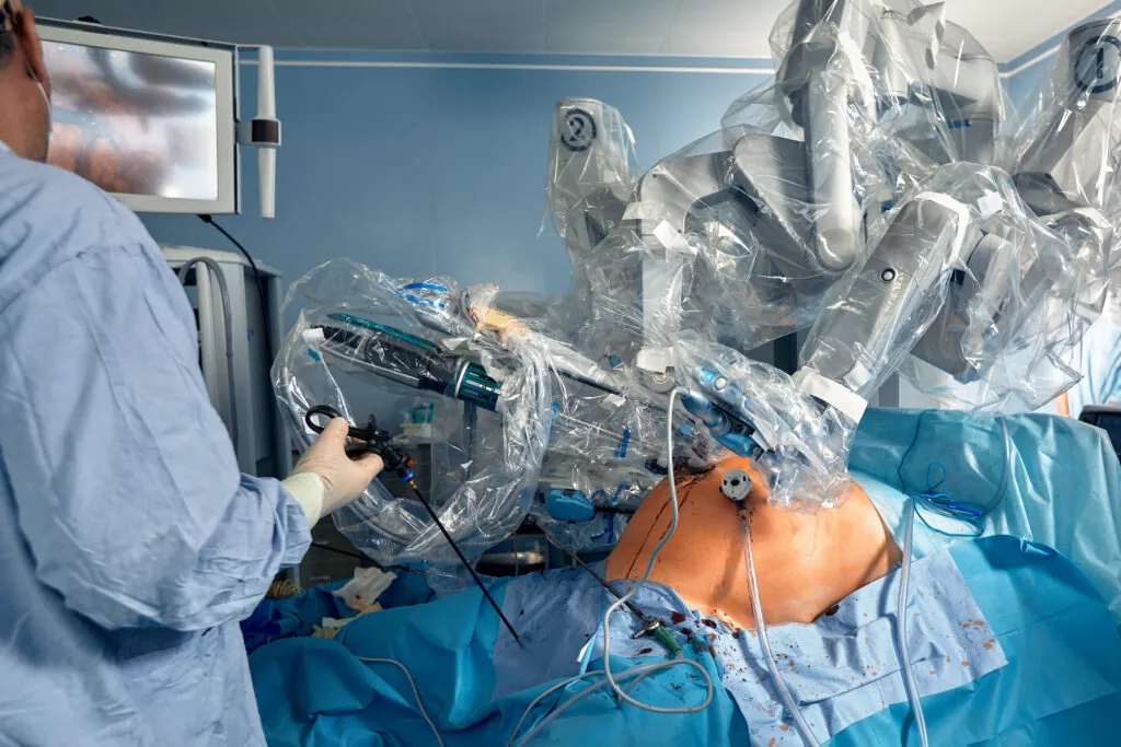
Surgical Options
Arthroscopic fat pad debridement aims to remove the portion of fat pad responsible for impingement while preserving functional tissue. The surgeon resects the scarred, hypertrophied posterior portion that enters the joint space during extension.
Surgery is typically reserved for patients who fail six months of comprehensive conservative treatment. Persistent symptoms despite appropriate physiotherapy, activity modification, and injection therapy indicate structural changes unlikely to resolve without intervention.
Post-operative rehabilitation emphasises early motion to prevent adhesion formation while protecting the resected fat pad from excessive stress. Full return to sport or demanding physical activities usually requires three to four months.
Outcomes following arthroscopic debridement are generally favourable, with many patients reporting substantial pain reduction. Incomplete resection or concurrent unrecognised pathology can be a cause of persistent symptoms following surgery.
From an Orthopaedic Perspective
Hoffa’s fat pad impingement often coexists with other anterior knee pathologies, including patellar tendinopathy and patellofemoral syndrome. A thorough clinical examination aims to identify all contributing factors—treating only the fat pad while missing concurrent problems may lead to incomplete symptom resolution.
Patients frequently respond well to conservative treatment when they commit to the necessary activity modifications and rehabilitation exercises. Patience during the initial inflammatory phase, and avoiding a return to aggravating activities before adequate healing, is important.
Managing Daily Activities
Avoid prolonged standing with knees locked in hyperextension—maintain a slight bend to reduce fat pad compression. When sitting for extended periods, periodically extend and flex the knee to prevent stiffness and maintain fat pad mobility.
Choose footwear with adequate cushioning to reduce impact transmission to the knee during walking. Avoid high heels, which shift body weight forward and increase extension loading on the knee.
Modify exercise routines to emphasise closed-chain strengthening in partial knee flexion ranges. Cycling, swimming, and elliptical training typically cause less fat pad irritation than running or jumping activities.
Use a pillow under the knee when sleeping on your back to maintain slight flexion. Avoid sleeping with legs fully straight, which sustains fat pad compression throughout the night.
✅ Quick Tip
When descending stairs, lead with your affected leg and keep your knee slightly bent throughout the step—this reduces the extension moment that pinches the fat pad compared to stepping down with a straight leg.
When to Seek Professional Help
- Anterior knee pain persisting beyond two weeks despite rest and ice
- Visible swelling below the kneecap that does not resolve
- Catching or blocking sensation when straightening the knee
- Pain preventing normal walking or stair climbing
- Symptoms following knee surgery that worsen rather than improve
- Night pain or symptoms accompanied by knee instability
Commonly Asked Questions
How long does Hoffa’s fat pad impingement take to heal?
Acute cases with mild inflammation often improve within four to six weeks of appropriate treatment. Chronic cases with established fibrosis may require several months of rehabilitation, and some patients need injection therapy or surgery. Healing time depends heavily on compliance with activity modification and consistent physiotherapy.
Can I continue exercising with fat pad impingement?
Exercise modification rather than complete rest is appropriate for most patients. Activities that avoid terminal knee extension—such as cycling with the seat slightly low or swimming with fins—maintain fitness without aggravating symptoms. High-impact and hyperextension activities should be avoided until symptoms resolve.
What is the difference between fat pad impingement and patellar tendinopathy?
Fat pad impingement typically produces pain below and around the kneecap, worsening with full extension and hyperextension. Patellar tendinopathy commonly causes pain at the inferior pole of the kneecap, typically aggravated by jumping and loading the tendon in flexion. Both conditions cause anterior knee pain but respond to different treatment approaches.
Will Hoffa’s fat pad impingement cause long-term knee damage?
Isolated fat pad impingement is not typically known to cause cartilage damage or arthritis. However, chronic inflammation can lead to permanent fat pad fibrosis that continues causing symptoms. Early appropriate treatment may help prevent these structural changes and improve long-term outcomes.
Is surgery always necessary for Hoffa’s fat pad impingement?
Many patients improve with conservative treatment including physiotherapy, activity modification, and anti-inflammatory measures. Surgery is reserved for cases that fail comprehensive non-operative management for at least six months.
Next Steps
Anterior knee pain below the kneecap that worsens with full extension, is associated with a catching or blocking sensation, and produces visible swelling around the patellar tendon are common indicators of Hoffa’s fat pad impingement. Conservative management, activity modification to avoid terminal extension loading, inflammation control, and targeted quadriceps and hip strengthening can resolve many cases without surgery. When these measures fail after six months, or when structural fat pad fibrosis is confirmed on MRI, arthroscopic debridement is a surgical option that typically yields favourable outcomes.
If you are experiencing anterior knee pain that worsens when straightening your leg, a catching or blocking sensation near full extension, or persistent swelling below the kneecap, our orthopaedic surgeon can evaluate your condition and discuss appropriate treatment options.