The kneecap (patella) relies on precise muscular balance to track smoothly within the femoral groove, and when that balance is disrupted, the result is patellofemoral pain syndrome, sometimes called runner’s knee.
This biomechanical problem causes friction and irritation on the cartilage underneath the patella, producing the characteristic aching pain around or behind the kneecap, and typically begins gradually before worsening over weeks or months.
Activities that require repeated knee bending, such as running, squatting, climbing stairs, or prolonged sitting, often intensify symptoms.
Recognising PFPS Symptoms
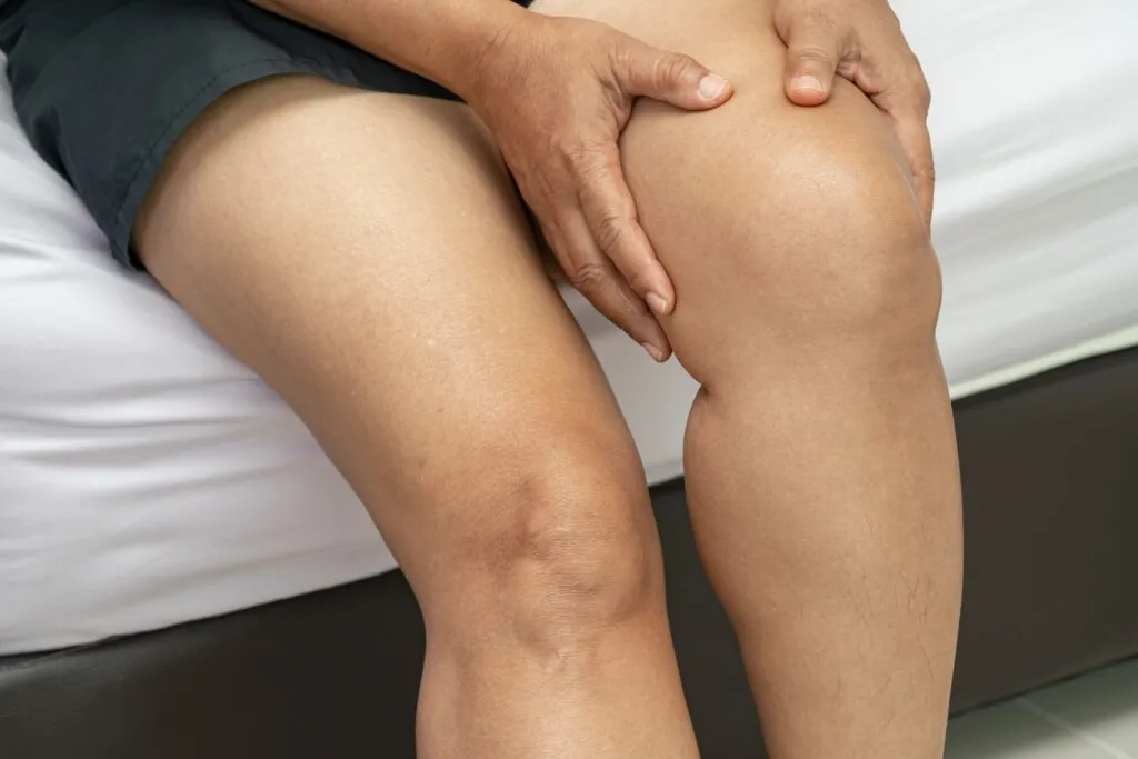
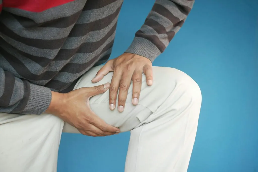
The hallmark symptom is a dull, aching pain located at the front of the knee, around the kneecap, or behind it. This pain typically increases with specific activities that load the patellofemoral joint.
Stair climbing and descent frequently provoke symptoms, with descending often causing more discomfort than ascending. The eccentric loading pattern during downward movement places significant stress on the patellofemoral joint. Squatting, kneeling, and lunging produce similar effects by compressing the kneecap against the femur.
“Cinema sign” or “theatre sign” describes pain that develops after prolonged sitting with knees bent. Maintaining knee flexion for extended periods sustains pressure on the patellofemoral joint, leading to stiffness and aching upon standing. Many people with PFPS instinctively extend their legs during prolonged sitting to relieve discomfort.
Grinding, clicking, or popping sensations (crepitus) may occur during knee movement. While crepitus itself isn’t always painful or concerning, its presence alongside pain suggests cartilage irritation. Some individuals notice mild swelling around the kneecap, though significant swelling is uncommon and may indicate a different condition.
Pain tends to worsen gradually with activity and improve with rest. However, complete inactivity can lead to stiffness and weakness that ultimately perpetuates the problem.
Understanding the Causes
Muscle Imbalances
The quadriceps muscle group controls patellar tracking through coordinated contraction of its four components. The vastus medialis obliquus (VMO), the teardrop-shaped muscle on the lower inner thigh just above the knee, plays a particularly important role in pulling the kneecap medially during movement.
When the VMO weakens relative to the outer quadriceps (vastus lateralis), the patella tends to drift laterally during knee extension. This lateral tracking concentrates forces on the outer edge of the patellofemoral joint. Hip muscle weakness, particularly in the gluteus medius and external rotators, compounds this problem by allowing the thigh to rotate inward during weight-bearing activities.
Tight structures on the lateral side of the knee, including the iliotibial band and lateral retinaculum, can further pull the patella off-centre. This combination of weak medial stabilisers and tight lateral structures creates the classic imbalance pattern seen in patellofemoral pain syndrome.
Structural Factors
Anatomical variations influence susceptibility to PFPS. A shallow femoral groove provides less guidance for patellar tracking. Patella alta (high-riding kneecap) reduces how deeply the patella sits within the groove during early knee flexion. A laterally positioned tibial tubercle, where the patellar tendon attaches below the knee, creates an outward-pulling force on the patella. These same structural factors are also relevant when the kneecap becomes unstable rather than simply painful.
Foot mechanics affect the entire lower limb chain. Excessive pronation (flat feet) causes internal rotation of the tibia and femur, altering patellofemoral joint alignment. Wide pelvis width relative to knee position increases the Q-angle, measured clinically as the angle formed between a line from the hip’s anterior superior iliac spine to the centre of the patella, and a line from the patella to the tibial tubercle, creating greater lateral force on the kneecap.
Activity-Related Factors
Sudden increases in training volume, intensity, or frequency commonly precipitate PFPS. The patellofemoral joint requires gradual adaptation to increased loading. Running mileage increases, adding hill training, or introducing new activities like jumping or court sports can overload unprepared tissues.
Training surfaces matter. Hard surfaces like concrete generate greater impact forces than softer alternatives. Cambered roads force asymmetrical loading on one knee. Worn or inappropriate footwear fails to provide adequate shock absorption or motion control.
Poor movement patterns during exercise, such as knees collapsing inward during squats, overstriding while running, or inadequate hip control during single-leg activities, repeatedly stress the patellofemoral joint in suboptimal positions.
Diagnostic Evaluation
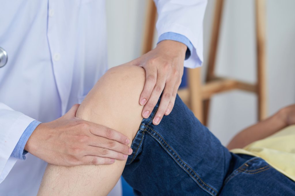
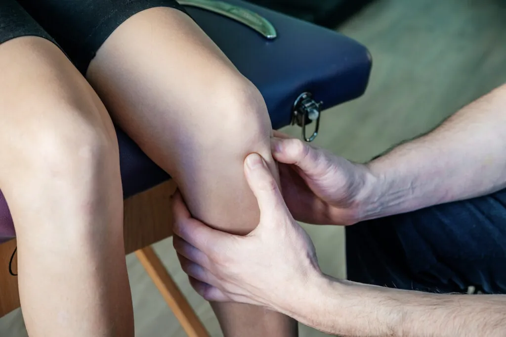
Clinical examination forms the foundation of PFPS diagnosis. An orthopaedic specialist will assess patellar tracking by observing knee extension against resistance, feeling for grinding beneath the kneecap, and testing patellar mobility. Provocative tests that reproduce symptoms help confirm the diagnosis.
Evaluation extends beyond the knee itself. Hip strength testing, particularly for abductors and external rotators, identifies proximal weakness contributing to poor leg alignment. Foot posture assessment reveals pronation patterns affecting tibial rotation. Flexibility testing of the quadriceps, hamstrings, iliotibial band, and calf muscles highlights tightness restricting normal movement.
Movement analysis during functional activities like squatting, stepping, or single-leg stance reveals dynamic alignment issues not apparent in static examination. Observing movements that provoke symptoms provides useful information about contributing factors.
Imaging isn’t routinely necessary for straightforward PFPS cases. Plain X-rays may be requested if structural abnormalities are suspected or if symptoms don’t respond to initial treatment. MRI is reserved for cases where cartilage damage, other soft tissue pathology, or alternative diagnoses need evaluation.
💡 Did You Know?
The patella is the largest sesamoid bone in the human body, a bone embedded within a tendon. It increases the mechanical advantage of the quadriceps muscle, allowing the thigh muscles to generate greater force during knee extension.
Treatment Approaches
Activity Modification
Initial management involves modifying activities that provoke symptoms rather than complete rest. Continuing pain-free movement maintains muscle strength and joint nutrition. Identifying specific triggers, perhaps running but not cycling, or descending stairs but not walking on flat ground, and temporarily reducing or avoiding those activities while maintaining others, may be helpful.
Cross-training with low-impact alternatives preserves cardiovascular fitness during recovery. Swimming, water running, cycling (with appropriate seat height adjustment), and elliptical training typically cause less patellofemoral stress than running or jumping activities.
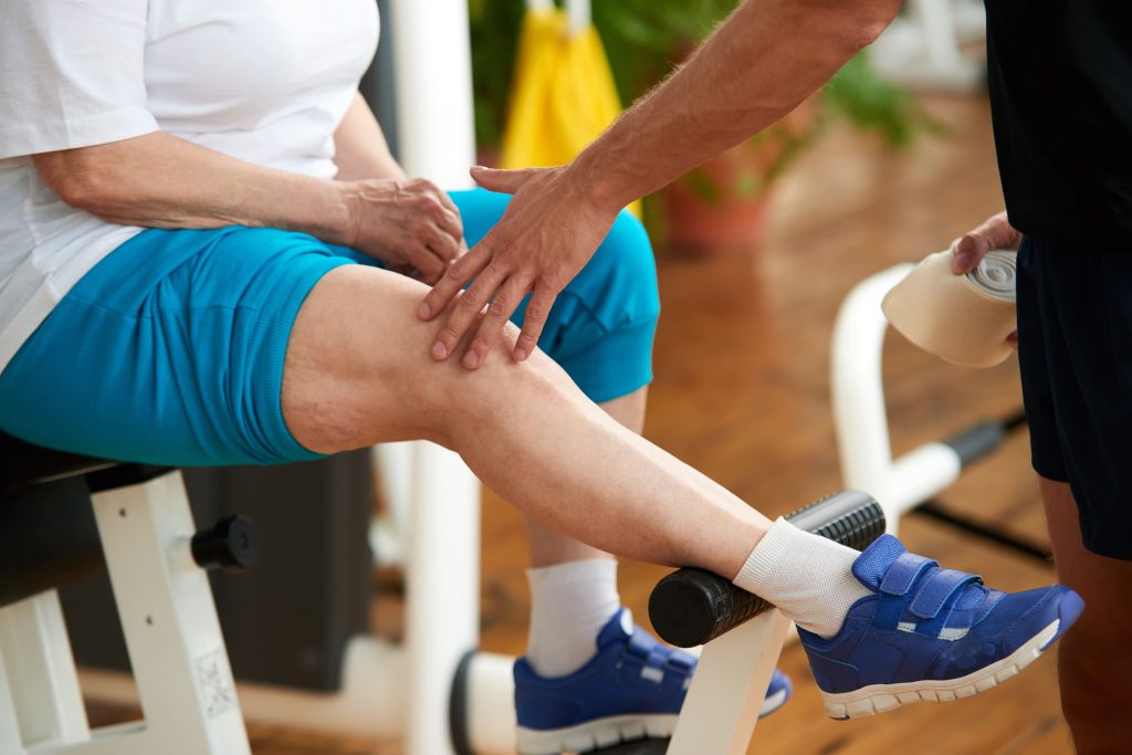
Rehabilitation Exercises
Targeted strengthening addresses the muscle imbalances underlying PFPS. VMO-focused exercises restore the medial quadriceps strength needed for proper patellar tracking. These include terminal knee extensions, partial-range squats, and step-downs with careful attention to knee alignment.
Hip strengthening is also important. Gluteal exercises, such as side-lying hip abduction, clamshells, bridges, and later single-leg activities, improve proximal control of leg alignment. When hip muscles adequately control femoral rotation, the knee remains better aligned during weight-bearing activities.
Stretching tight structures restores flexibility balance. Quadriceps, hamstring, iliotibial band, and calf stretches reduce tension that restricts normal movement. Foam rolling provides additional soft tissue mobilisation.
Neuromuscular training teaches proper movement patterns. Practising squats, lunges, and step-downs in front of a mirror while maintaining knee alignment over the foot retrains motor patterns. Single-leg balance exercises challenge dynamic stability.
Progression follows a logical sequence: pain-free range of motion, then strengthening in stable positions, advancing to dynamic exercises, and finally returning to sport-specific activities. Rushing this progression risks symptom recurrence.
Additional Interventions
Patellar taping provides temporary symptom relief by altering patellar position or reducing abnormal stress. McConnell’s taping techniques aim to medially glide and tilt the patella. While taping doesn’t address underlying causes, it can enable pain-free exercise during rehabilitation.
Knee braces designed for patellofemoral conditions incorporate patellar stabilisation features. These may help during activity while muscle strength develops, though they shouldn’t replace rehabilitation exercises.
Foot orthoses (custom or prefabricated insoles) address excessive pronation and its effects on tibial and femoral rotation. Proper footwear assessment ensures appropriate support and cushioning for the foot type and activities involved.
Manual therapy, including soft tissue massage, joint mobilisation, and dry needling, may complement exercise-based treatment. These techniques address local tissue restrictions and can reduce pain, facilitating more effective strengthening exercises.
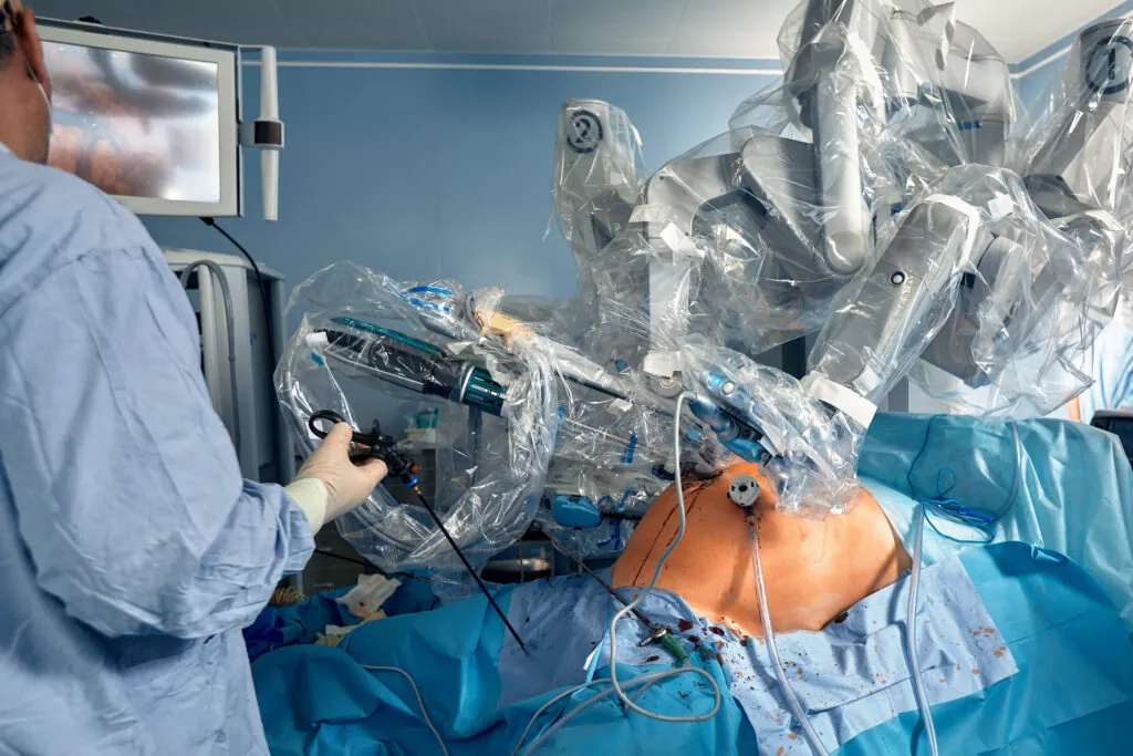
When Surgery Becomes an Option
Surgical intervention is rarely necessary for PFPS and is considered only after several months of comprehensive rehabilitation have failed to provide adequate relief. When surgery is indicated, treatment options include:
- Lateral release divides the tight lateral retinaculum, reducing its pull on the patella. This procedure addresses structural tightness but doesn’t correct muscle weakness.
- Tibial tubercle transfer repositions the patellar tendon attachment, changing the line of pull on the patella. This more involved procedure addresses significant malalignment not correctable through rehabilitation alone.
- Cartilage procedures may be appropriate if significant cartilage damage has occurred. Options range from smoothing damaged cartilage (chondroplasty) to various cartilage restoration techniques.
Surgery addresses structural problems but doesn’t replace the need for rehabilitation. Post-operative strengthening remains important for recovery.
Preventing Recurrence
Long-term management focuses on maintaining the strength and flexibility gains achieved during rehabilitation. Continuing hip and quadriceps strengthening exercises at a reduced frequency prevents the gradual weakening that allows symptoms to return.
Training modifications reduce reinjury risk. Gradual progression of activity intensity and volume allows tissue adaptation. Incorporating rest days prevents cumulative overload. Varying training surfaces and activities distribute stress across different structures.
Movement awareness during daily activities and exercise helps maintain proper alignment. Monitoring for early warning signs, such as mild aching after activity or stiffness after prolonged sitting, allows prompt intervention before symptoms escalate.
⚠️ Important Note
Pushing through increasing knee pain during exercise can worsen cartilage irritation and prolong recovery. Mild discomfort that doesn’t worsen during activity and settles quickly afterwards is generally acceptable, but pain that intensifies or persists signals the need to modify your approach.
When to Seek Professional Help
- Knee pain persists beyond several weeks despite rest and activity modification
- Pain severe enough to limit daily activities or disturb sleep
- Knee giving way or feeling unstable during movement
- Significant swelling, warmth, or redness around the knee
- Locking or catching sensations are preventing full knee movement
- Pain following a specific injury or trauma to the knee
- Symptoms not improving after several weeks of self-directed exercises
Commonly Asked Questions
How long does it take for patellofemoral pain syndrome to heal?
Recovery timeframes vary based on symptom severity, contributing factors, and rehabilitation compliance. Mild cases may improve within several weeks of appropriate exercise and activity modification. More established cases typically require several months of consistent rehabilitation. Factors like significant muscle weakness or structural malalignment extend recovery timelines.
Can I continue running with PFPS?
This depends on symptom severity. If running causes only mild discomfort that doesn’t worsen during the run and settles quickly afterwards, continuing at reduced volume may be acceptable. Significant pain during or after running indicates the need for temporary cessation. Many runners successfully return to full training after addressing underlying biomechanical issues.
Is patellofemoral pain syndrome the same as chondromalacia patellae?
These terms are related but distinct. Chondromalacia patellae specifically refers to softening and damage of the cartilage on the undersurface of the patella. PFPS is a broader clinical diagnosis based on symptoms, which may or may not involve visible cartilage changes. Many patients with PFPS have normal cartilage, while some cartilage changes seen on imaging cause no symptoms.
Will PFPS lead to arthritis?
The relationship between PFPS and future osteoarthritis remains unclear. Cartilage irritation doesn’t inevitably progress to arthritis, particularly when contributing factors are addressed. Maintaining good muscle strength, healthy body weight, and appropriate activity levels supports long-term joint health.
Are there exercises I should avoid with PFPS?
Deep squats, lunges beyond comfortable range, and high-impact jumping activities often aggravate symptoms and are best avoided initially. Leg extension machines with heavy resistance at full range can overload the patellofemoral joint. As rehabilitation progresses and symptoms improve, many of these activities can be carefully reintroduced with proper technique.
Next Steps
PFPS typically responds well when the specific contributing factors, muscle imbalances, flexibility deficits, and movement patterns, are accurately identified and addressed through targeted rehabilitation. Structured rehabilitation forms the foundation of long-term recovery. Surgical intervention is rarely required but remains an option after conservative pathways have been fully explored.
If you are experiencing anterior knee pain, clicking or grinding sensations around the kneecap, or discomfort with stair descent and prolonged sitting, our orthopaedic surgeon can evaluate your patellofemoral alignment and recommend treatment based on your specific biomechanical findings.